 Back
6A-LE3 - Anatomy & Physiology
 3:56

Terms in this set (20)
Skeletal cartilage is made of highly resilient cartilage tissue that consists primarily of water and contains no blood vessels or nerves.
The perichondrium is a dense connective tissue layer surrounding cartilage that resists outward expansion and provides blood vessels for nutrient delivery.
- Hyaline cartilage: Most abundant, contains only collagen fibers.
- Elastic cartilage: Contains elastic fibers, found in external ear and epiglottis.
- Fibrocartilage: Thick collagen fibers, great tensile strength, found in menisci and vertebral discs.
- Appositional growth: New matrix laid on cartilage surface, increasing length.
- Interstitial growth: Chondrocytes divide within lacunae, secreting new matrix, increasing thickness.
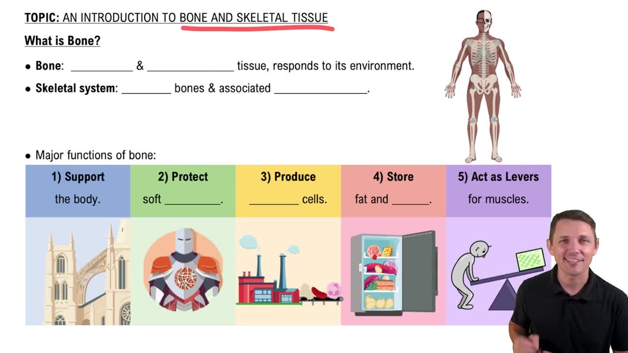
- Support
- Protection
- Movement
- Mineral and growth factor storage
- Blood cell formation (hematopoiesis)
- Triglyceride (fat) storage
- Hormone production (osteocalcin)
Divided into axial skeleton (skull, vertebral column, rib cage) and appendicular skeleton (limbs and girdles).
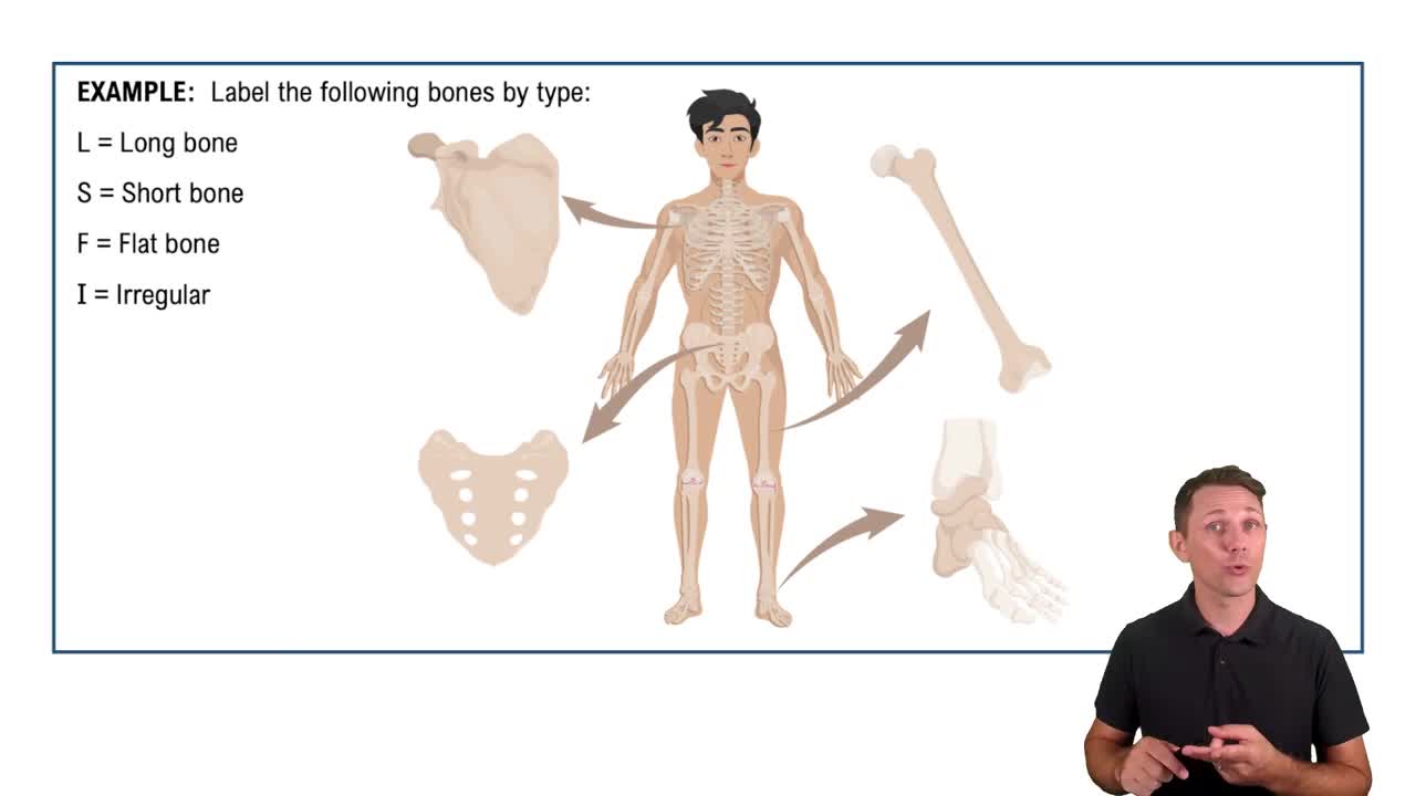
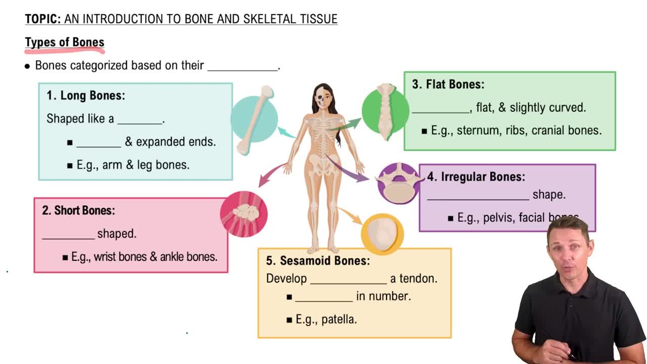
- Long bones: Longer than wide, limb bones.
- Short bones: Cube-shaped, e.g., wrist and ankle.
- Flat bones: Thin, flat, slightly curved, e.g., sternum.
- Irregular bones: Complicated shapes, e.g., vertebrae.
Compact bone: Dense outer layer, smooth and solid.
Spongy bone: Honeycomb of trabeculae with spaces filled with marrow.
- Diaphysis: Shaft with compact bone surrounding medullary cavity.
- Epiphyses: Ends with compact bone outside, spongy inside, covered by articular cartilage.
- Epiphyseal line: Remnant of growth plate.
- Periosteum: Double-layered membrane covering external bone surfaces except joints; contains osteogenic cells, nerves, and blood vessels.
- Endosteum: Delicate membrane covering internal bone surfaces and trabeculae; contains osteogenic cells.
Red marrow is found in trabecular cavities of spongy bone and flat bone diploë; it is the site of hematopoiesis (blood cell formation).
- Projections: Sites of muscle and ligament attachment.
- Depressions: Grooves or indentations for vessels or joints.
- Openings: Holes or canals for blood vessels and nerves.
Osteogenic cells are mitotically active stem cells in periosteum and endosteum that differentiate into osteoblasts or bone-lining cells.
Osteoblasts are bone-forming cells that secrete unmineralized bone matrix called osteoid.
Osteocytes are mature bone cells in lacunae that maintain bone matrix and act as stress/strain sensors.
Osteoclasts are large multinucleate cells derived from hematopoietic stem cells that resorb bone.
An osteon is the structural unit of compact bone, consisting of concentric lamellae around a central (Haversian) canal containing blood vessels and nerves.
Canaliculi are tiny canals connecting lacunae and the central canal, allowing nutrient and waste exchange between osteocytes.
- Organic components: Cells and osteoid (collagen and ground substance) providing flexibility.
- Inorganic components: Hydroxyapatites (calcium phosphate crystals) providing hardness and resistance to compression.
Bone collagen fibers run in different directions in lamellae, and sacrificial bonds in collagen dissipate energy to prevent fractures.