Articulation between the mandible and temporal bone; acts as a modified hinge and gliding joint with moderate stability.
Shoulder Joint
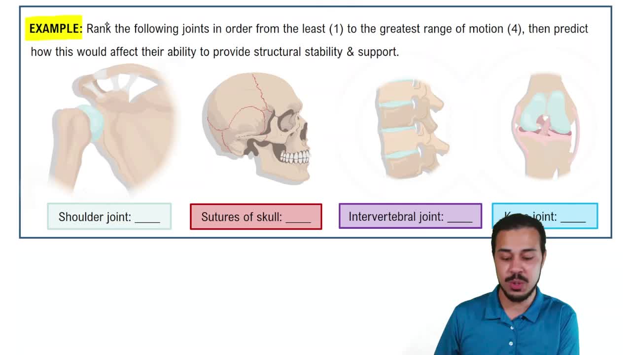
Articulation between the humerus and scapula; ball-and-socket joint with low stability and high mobility.
Elbow Joint
Articulation between the humerus, radius, and ulna; stable uniaxial hinge joint allowing flexion and extension.
Hip Joint
Articulation between the femur and pelvis; ball-and-socket joint with high stability due to a deep socket and strong ligaments.
Knee Joint
Articulation between the femur, tibia, and patella; modified hinge joint allowing flexion, extension, and slight rotation; stability from ligaments and menisci.
Articular Surfaces of Temporomandibular Joint
Egg-shaped condylar process of mandible articulates with mandibular fossa and articular tubercle of temporal bone.
Function of Articular Disc in Temporomandibular Joint
Divides synovial cavity into two compartments: inferior for hinge-like elevation/depression, superior for anterior gliding and lateral excursion.
Common Temporomandibular Joint Dislocation
Dislocation occurs anteriorly into infratemporal fossa, leaving mouth wide open; realignment requires pushing mandible inferiorly and posteriorly.
Shoulder Joint Articular Architecture
Multiaxial ball-and-socket formed by humerus head and shallow glenoid cavity, deepened by fibrocartilaginous glenoid labrum; enclosed by thin, loose capsule.
Coracohumeral Ligament
Primary strong ligament supporting the weight of the upper limb in the shoulder joint.
Role of Biceps Brachii Tendon (Long Head) in Shoulder
Acts as a 'superstabilizer' securing the humerus head within the glenoid cavity.
Rotator Cuff Muscles
Four muscle tendons encircling shoulder joint: subscapularis, supraspinatus, infraspinatus, and teres minor.
Common Shoulder Dislocation Direction
Most vulnerable anteriorly and inferiorly; dislocations occur forward and downward when humerus is abducted and rotated laterally.
Elbow Joint Articular Mechanism
Uniaxial hinge permitting flexion and extension; trochlear notch of ulna grips trochlea of humerus.
Anular Ligament Function
Surrounds head of radius, allowing rotation during pronation and supination without affecting hinge movement.
 Back
 04:06