Skip to main content
Anatomy & Physiology
My Course
Learn
Exam Prep
AI Tutor
Study Guides
Textbook Solutions
Flashcards
Explore
Try the app
My Course
Learn
Exam Prep
AI Tutor
Study Guides
Textbook Solutions
Flashcards
Explore
Try the app
Back
Anatomy & Physiology: Blood and Hemostasis
You can tap to flip the card.
Primary functions of blood
You can tap to flip the card.
👆
Primary functions of blood
Transport
of O2, nutrients, wastes, and hormones;
Regulation
of body temperature, pH, and fluid volume;
Protection
against blood loss and infection.
Track progress
Control buttons has been changed to "navigation" mode.
1/25
Recommended videos
Guided course
2:36
Introduction to Blood Example 1
10935
views
327
rank
Guided course
7:49
Composition of Blood
11534
views
330
rank
Guided course
1:55
Introduction to Blood Example 2
9049
views
219
rank
Terms in this set (25)
Hide definitions
Primary functions of blood
Transport
of O2, nutrients, wastes, and hormones;
Regulation
of body temperature, pH, and fluid volume;
Protection
against blood loss and infection.
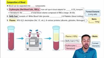
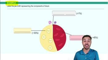
Composition of blood
Blood consists of
plasma
(55%),
erythrocytes
(45%), and a thin
buffy coat
of leukocytes and platelets (<1%).
Erythrocyte structure
Biconcave, anucleate cells filled with hemoglobin; flexible due to spectrin protein; optimized for gas transport.
Hemoglobin structure and function
Consists of four polypeptide chains (two alpha, two beta) each with a heme group containing iron that binds O2 reversibly.
Erythropoiesis
Process of RBC formation in red bone marrow, taking about 15 days, regulated by erythropoietin and requiring iron, B12, and folic acid.
Regulation of erythropoiesis
Stimulated by hypoxia via kidney-released erythropoietin; balanced to avoid hypoxia or excessive blood viscosity.
Life span and fate of erythrocytes
Live 100-120 days; old RBCs are phagocytized in spleen; hemoglobin components recycled or excreted as bilirubin.
Anemia
Condition with abnormally low O2-carrying capacity due to blood loss, decreased RBC production, or increased RBC destruction.
Polycythemia
Excess RBCs increasing blood viscosity; caused by bone marrow cancer or secondary to hypoxia; blood doping mimics this.
Leukocytes (WBCs) characteristics
Complete cells with nuclei; defend against disease; can exit bloodstream via diapedesis; two groups: granulocytes and agranulocytes.
Granulocytes vs Agranulocytes
Granulocytes have visible cytoplasmic granules (neutrophils, eosinophils, basophils); agranulocytes lack granules (lymphocytes, monocytes).
Leukopoiesis
WBC production stimulated by interleukins and colony-stimulating factors; originates from hemocytoblast stem cells.
Platelets origin and function
Cytoplasmic fragments of megakaryocytes; contain clotting chemicals; form temporary plugs to seal vessel breaks.
Hemostasis steps
1) Vascular spasm (vasoconstriction), 2) Platelet plug formation, 3) Coagulation (fibrin mesh formation).
Platelet plug formation
Platelets adhere to exposed collagen, release ADP, serotonin, and thromboxane A2, promoting aggregation and vascular spasm.
Coagulation pathways
Intrinsic (clotting factors in blood) and extrinsic (tissue factor outside blood) pathways activate factor X, leading to thrombin formation.
Common coagulation pathway
Thrombin converts fibrinogen to fibrin, forming a mesh that stabilizes the clot; factor XIII cross-links fibrin.
Clot retraction and fibrinolysis
Platelets contract to shrink clot; PDGF and VEGF promote vessel repair; plasmin dissolves clot after healing.
Factors limiting clot growth
Removal/dilution of clotting factors; antithrombin III and heparin inhibit thrombin; endothelial cells secrete anticoagulants.
ABO blood group system
Based on presence of A and/or B antigens on RBCs; plasma contains antibodies against absent antigens; determines compatibility.
Rh blood group system
Presence (Rh+) or absence (Rh−) of D antigen; Rh− individuals form anti-Rh antibodies only after exposure to Rh+ blood.
Hemolytic disease of the newborn
Occurs when Rh− mother sensitized to Rh+ fetus blood produces antibodies that destroy fetal RBCs in subsequent pregnancies.
Transfusion reactions
Mismatched blood causes agglutination, hemolysis, reduced oxygen capacity, and kidney damage; symptoms include fever and chills.
Universal donor and recipient blood types
Type O is universal donor (no A/B antigens); Type AB is universal recipient (no anti-A/B antibodies).
Blood typing and cross matching
Mix donor RBCs with recipient serum and vice versa to detect agglutination; ensures compatibility before transfusion.
 BackBack
 2:36
2:36