 Back
Anatomy & Physiology: Blood, Heart, and Blood Vessels
 2:36

Terms in this set (30)
Transport of gases, nutrients, and wastes; regulation of pH, temperature, and water content; protection against blood loss and infection.
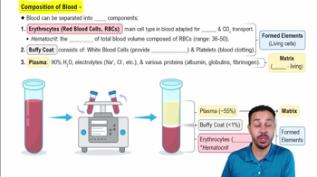
Viscous, sticky fluid with a temperature of about 38°C, pH 7.35-7.45, and volume of 4-6 liters in adults.
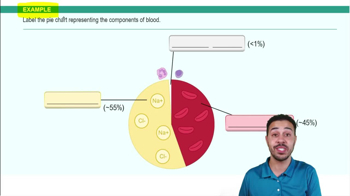
Plasma (55%) and formed elements: red blood cells (RBCs), white blood cells (WBCs), and platelets.
Hematopoiesis occurs in red bone marrow producing RBCs, WBCs, and platelets from hematopoietic stem cells.
Transport oxygen from lungs to tissues and carbon dioxide from tissues to lungs using hemoglobin.
Defend the body against pathogens and remove debris through immune responses.
Participate in hemostasis by forming platelet plugs and releasing chemicals for blood clotting.
1. Vascular spasm constricts blood vessels.
2. Platelet plug formation seals small breaks.
3. Blood clotting stabilizes the plug with fibrin.
Triggered by external trauma causing blood to escape vessels; involves tissue factor (factor III) activating factor VII.
Activated by damage inside the blood vessel; involves factors XII, XI, IX, and VIII.
Converges from intrinsic and extrinsic pathways; activates factor X leading to conversion of prothrombin to thrombin and fibrinogen to fibrin.
Based on presence of antigens (A, B) on RBCs and antibodies in plasma; includes ABO and Rh systems.
A condition with reduced oxygen-carrying capacity due to low RBC count or hemoglobin abnormalities.
Genetic disorder causing abnormal hemoglobin that distorts RBCs into a sickle shape, leading to blockages and anemia.
Epicardium (outer), myocardium (muscle layer), and endocardium (inner lining).
Four chambers: two atria (upper) and two ventricles (lower) separated by septa.
Atrioventricular valves (tricuspid and mitral) and semilunar valves (pulmonary and aortic) prevent backflow.
Blood supply to the heart muscle via coronary arteries and veins.
Includes sinoatrial (SA) node, atrioventricular (AV) node, bundle of His, bundle branches, and Purkinje fibers to coordinate heartbeat.
P wave: atrial depolarization.
QRS complex: ventricular depolarization.
T wave: ventricular repolarization.
Includes atrial systole, ventricular systole, and diastole with pressure and volume changes in chambers.
First sound (S1) is closure of AV valves; second sound (S2) is closure of semilunar valves.
Volume of blood pumped per minute; calculated as \(\text{CO} = \text{HR} \times \text{SV}\).
Influenced by preload, contractility, and afterload.
Controlled by autonomic nervous system and hormones like epinephrine.
Three layers: tunica intima, tunica media, and tunica externa.
Arteries, arterioles, capillaries, venules, and veins with distinct functions and structures.
Movement of gases, nutrients, and wastes between blood and tissues via diffusion, filtration, and osmosis.
Blood pressure, vascular resistance, blood viscosity, vessel diameter, and length.
Controlled by cardiovascular center in brainstem with short-term (neural/hormonal) and long-term (renal) mechanisms.