 Back
Anatomy & Physiology: Bone and Skeletal System Fundamentals
 3:56

Terms in this set (28)
Tough connective-tissue covering on the outside of bone except where articular cartilage covers joints; contains blood vessels, nerves, and bone-forming cells.
Thin membrane lining internal bone surfaces, including the medullary cavity and trabeculae.
Cylindrical structural unit of compact bone; concentric rings of bone matrix surround a central canal containing blood vessels and nerves.
Mature bone cell housed in lacunae that maintains bone tissue.
Bone-forming cell that secretes bone matrix (osteoid).
Cell that breaks down or resorbs bone tissue during remodeling.
Dense, strong bone tissue organized into osteons; provides strength and resistance to bending.
Bone tissue arranged as trabeculae with spaces containing marrow; lighter and aligns with stress.
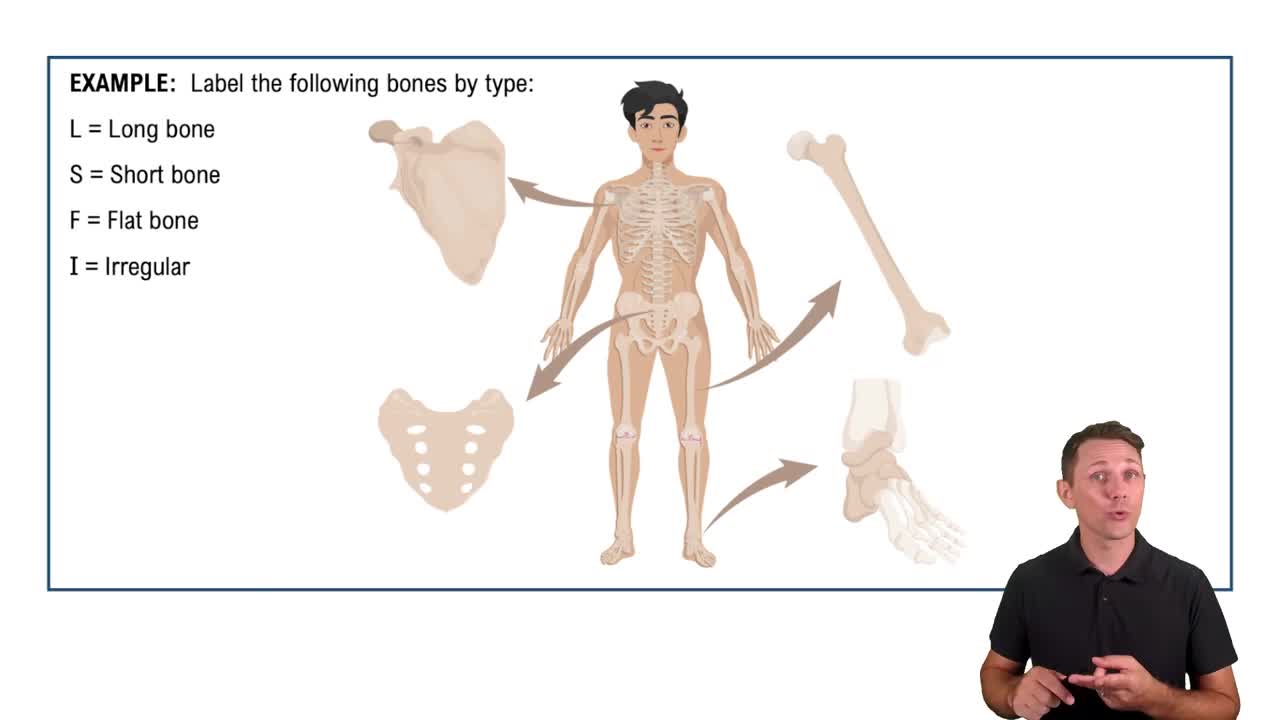
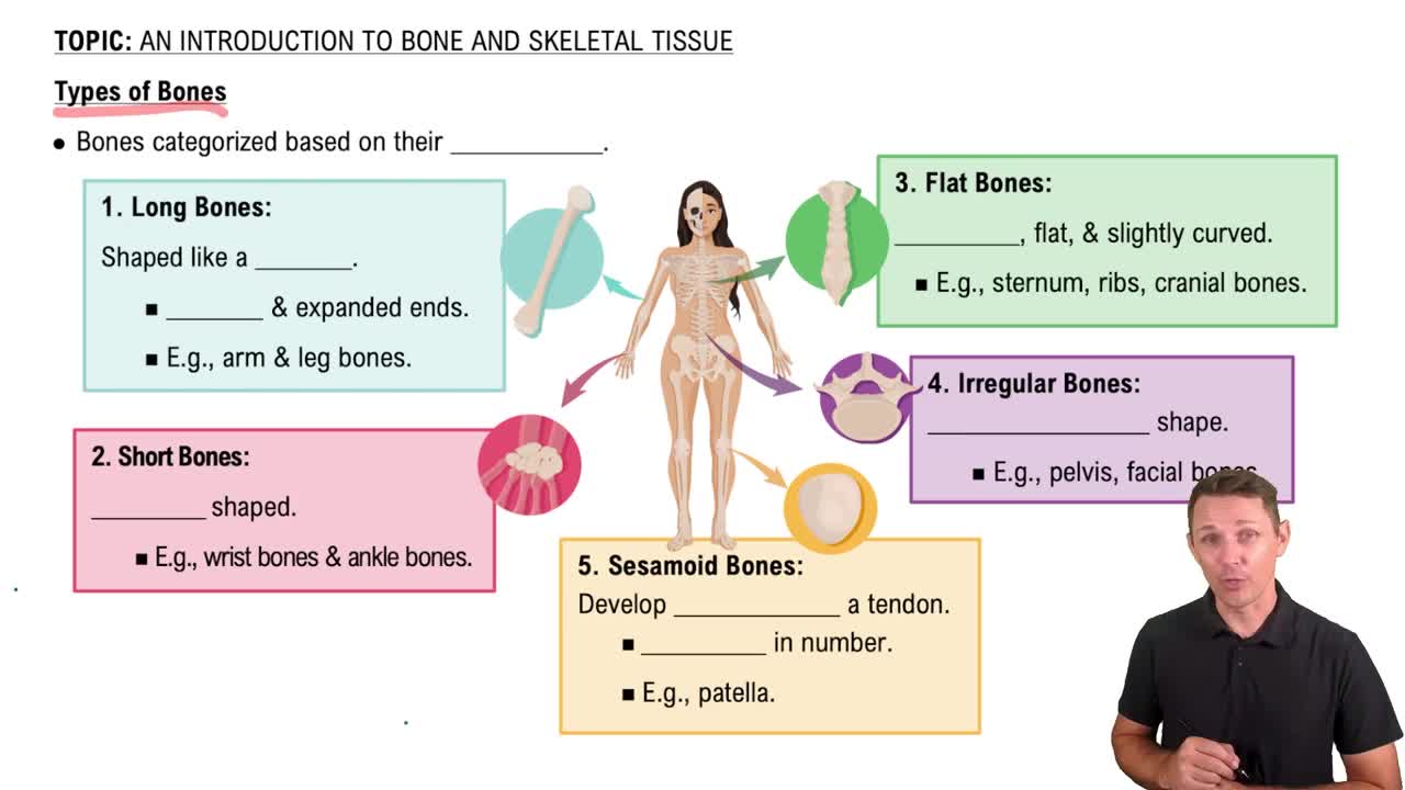
Bones longer than wide with a shaft and two ends; examples include femur and humerus.
Bone forms directly within connective tissue membrane without a cartilage model; forms flat skull bones and clavicle.
Bone replaces a hyaline cartilage model; forms most bones including long bones.
Cartilage growth plate in long bones where lengthening occurs; later becomes the epiphyseal line.
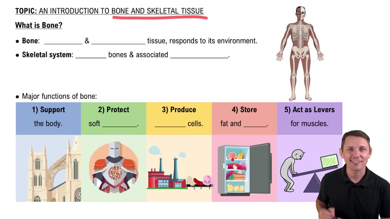
Support, protection, movement, mineral storage/release, and blood-cell formation (hematopoiesis).
Continuous process where osteoclasts resorb bone and osteoblasts deposit new matrix to repair damage and regulate minerals.
Hormone that raises blood calcium by increasing bone resorption, kidney calcium retention, and vitamin D activation.
Hormone from thyroid C cells that lowers blood calcium by inhibiting osteoclast activity; less dominant than PTH in adults.
Central framework including skull, vertebral column, ribs, and sternum; consists of 80 bones.
Includes upper and lower limbs plus pectoral and pelvic girdles; consists of 126 bones.
Cervical and lumbar curves are convex forward (secondary); thoracic and sacral curves are convex backward (primary).
Abnormal lateral (side-to-side) curvature of the vertebral column.
Clavicle and scapula; attaches upper limbs, prioritizes mobility, and is relatively light.
Right and left hip bones; attaches lower limbs, transfers body weight, and prioritizes stability and organ protection.
Wider and shallower with larger pelvic inlet/outlet and wider subpubic angle to accommodate childbirth.
Narrower and deeper with smaller pelvic inlet/outlet and narrower subpubic angle; generally more robust bone markings.
Hematoma formation, soft callus formation, bony callus formation, and remodeling into lamellar bone.
Tiny channels connecting lacunae that allow osteocytes to exchange nutrients and wastes.
Hollow space in the diaphysis of a long bone; contains yellow marrow in adults.
Bone that develops within a tendon to reduce friction or alter tendon pull; the patella is the largest example.