 Back
Anatomy & Physiology: Bone, Skeletal System, Joints, and Nervous System Study Guide
 3:56

Terms in this set (62)
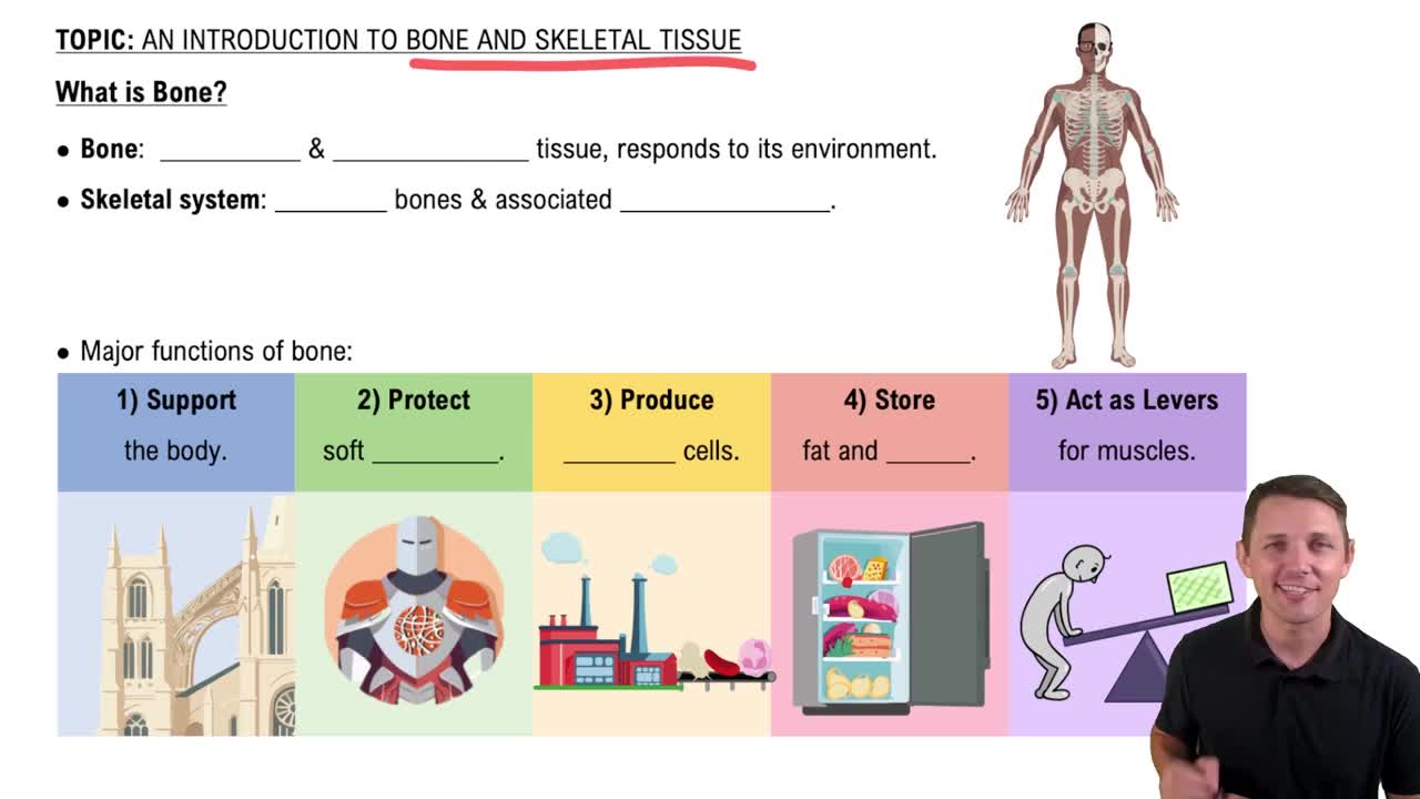
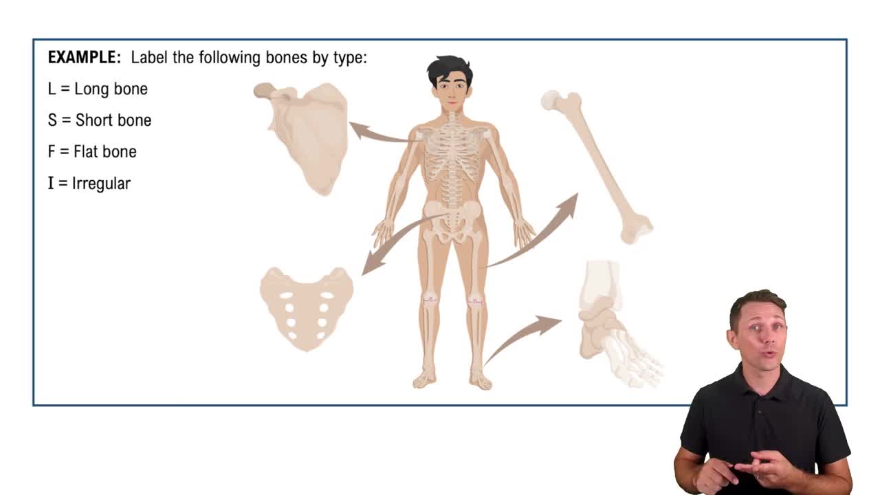
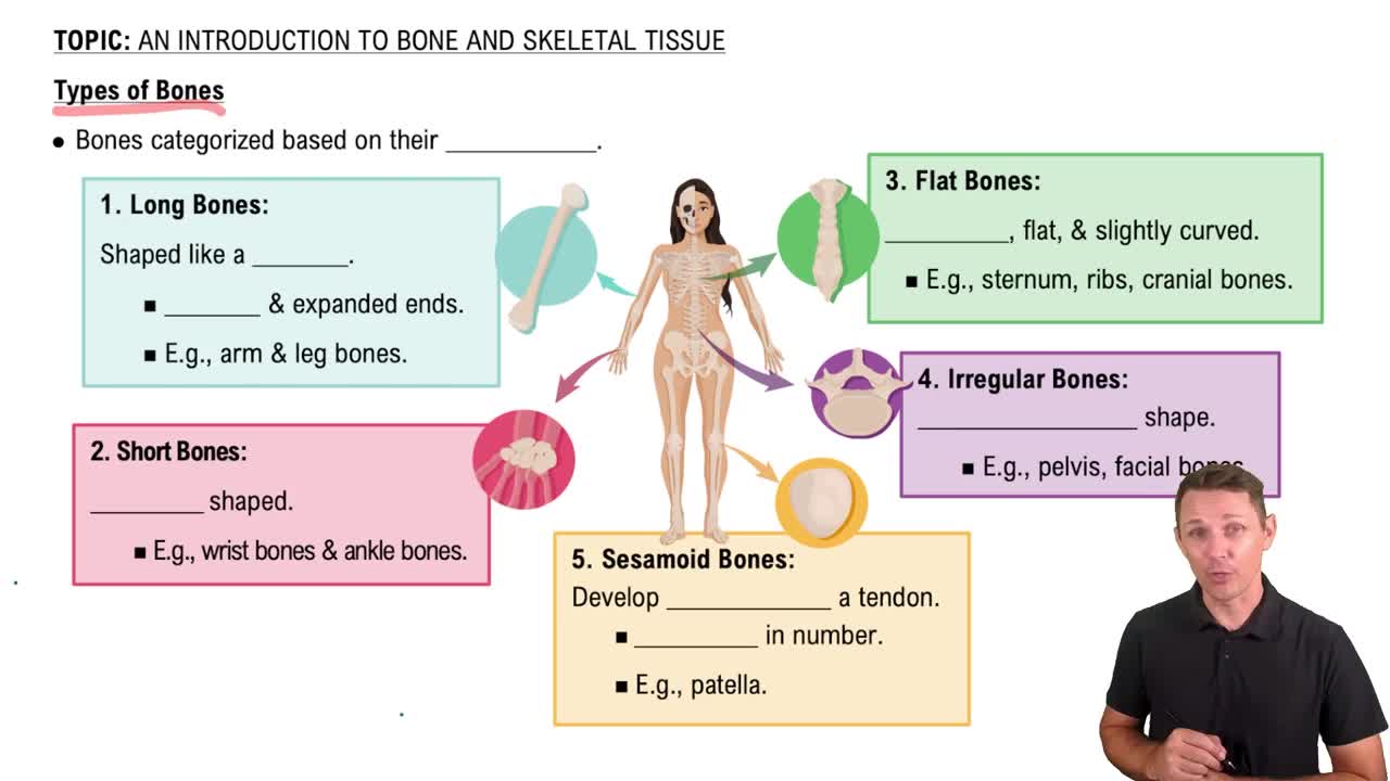
Long, short, flat, and irregular bones differ in shape and function. Long bones are longer than wide, short bones are cube-shaped, flat bones are thin and curved, and irregular bones have complex shapes.
Osteoblasts build bone, osteocytes maintain bone, osteoclasts resorb bone, and bone lining cells regulate calcium and phosphate.
A bone with a diaphysis (shaft) and epiphyses (ends) is a long bone.
Collagen deficiency weakens bone matrix, making bones brittle and prone to fractures, as seen in osteogenesis imperfecta.
Osteoblasts synthesize and deposit bone matrix; osteoclasts break down bone tissue during resorption.
An osteon includes central canal (blood vessels), lamellae (concentric bone layers), lacunae (spaces housing osteocytes), and canaliculi (channels for cell communication).
Interstitial growth involves chondrocyte division, matrix secretion, calcification, and replacement by bone tissue at the epiphyseal plate.
Bone forms directly from mesenchymal tissue without a cartilage model, mainly in flat bones of the skull.
Osteoblasts differentiate from mesenchymal cells and secrete bone matrix to form bone tissue.
Bone develops by replacing a hyaline cartilage model, typical of long bones.
Intramembranous ossification forms bone directly from mesenchyme; endochondral ossification replaces cartilage with bone.
The epiphyseal plate is a cartilage growth plate in children; the epiphyseal line is the ossified remnant in adults.
Increased testosterone and estrogen during puberty stimulate growth plate activity; estrogen causes earlier plate closure, explaining why women are generally shorter.
A person who exercises experiences more bone deposition due to mechanical stress stimulating osteoblast activity.
Parathyroid hormone increases blood calcium by stimulating osteoclasts; calcitriol enhances calcium absorption in the intestines.
Vitamin C deficiency impairs collagen synthesis weakening bone; vitamin D deficiency reduces calcium absorption causing weak, soft bones.
Ossification is bone tissue formation; calcification is deposition of calcium salts in tissue, which can occur in ossification or other tissues.
Fracture repair includes hematoma formation, fibrocartilaginous callus formation, bony callus formation, and bone remodeling.
The epiphyseal plate allows lengthwise bone growth by cartilage proliferation and ossification; damage can stunt growth.
No, calcification of cartilage matrix is necessary before ossification during longitudinal growth.
Bone resorption exceeds deposition, leading to bone loss and weakened bones.
Axial skeleton includes skull, vertebrae, ribs; appendicular skeleton includes limbs and girdles.
The pelvic girdle consists of the two hip bones (ilium, ischium, pubis) and the sacrum.
The pectoral girdle includes the clavicle and scapula.
Cranial bones protect the brain; facial bones form the face structure.
Upper limb: humerus, radius, ulna, carpals, metacarpals, phalanges. Lower limb: femur, tibia, fibula, tarsals, metatarsals, phalanges.
Synarthrosis: immovable (skull sutures), amphiarthrosis: slightly movable (intervertebral discs), diarthrosis: freely movable (shoulder joint).
Fibrous joints are connected by dense connective tissue and allow little or no movement.
All fibrous joints lack a joint cavity and are connected by dense fibrous connective tissue.
Syndesmoses are fibrous joints with bones connected by ligaments, allowing slight movement.
Cartilaginous joints connect bones with cartilage and allow limited movement.
Synchondroses (hyaline cartilage, e.g., epiphyseal plate), symphyses (fibrocartilage, e.g., pubic symphysis).
Cartilage acts as a shock absorber and allows slight movement between vertebrae.
Synovial joints have a fluid-filled cavity allowing free movement; fibrous and cartilaginous joints do not.
Nonaxial (gliding, carpals), uniaxial (hinge, elbow), biaxial (condyloid, knuckle), multiaxial (ball-and-socket, shoulder).
Synovial fluid lubricates joints, nourishes cartilage, and absorbs shock.
Hinge joints are more stable due to limited movement (elbow); ball-and-socket joints allow more movement but less stability (shoulder).
Elbow has stronger ligament support and less complex movement than the knee, making it more stable.
Anterior cruciate ligament, posterior cruciate ligament, medial collateral ligament, lateral collateral ligament.
Both are ball-and-socket joints allowing multiaxial movement and have a joint capsule with synovial fluid.
Damage leads to pain, reduced mobility, and arthritis due to loss of smooth joint surface.
High mobility and shallow socket make the shoulder joint prone to dislocation and injury.
Movements like supination/pronation (forearm), inversion/eversion (foot), and opposition (thumb) occur at specific joints.
Cartilage degeneration, decreased synovial fluid, and osteoarthritis causing pain and stiffness.
Oligodendrocytes myelinate CNS axons; Schwann cells myelinate PNS axons.
Divided into CNS (brain and spinal cord) and PNS (nerves and ganglia).
High K+ inside, high Na+ outside; resting potential maintained by Na+/K+ pumps and leak channels.
Myelination increases conduction speed by enabling saltatory conduction between nodes of Ranvier.
Leak channels are always open; gated channels open or close in response to stimuli.
Ion concentration gradients and selective membrane permeability to K+.
Depolarization to threshold opens Na+ channels, repolarization opens K+ channels, hyperpolarization follows before returning to rest.
Toxin blocks Na+ channels, preventing action potentials and nerve signal transmission.
Na+ channels are inactivated and cannot reopen immediately after an action potential.
Ca2+ influx triggers neurotransmitter vesicle fusion and release into synaptic cleft.
Chemical synapses use neurotransmitters; electrical synapses use gap junctions for direct ion flow.
EPSP (excitatory postsynaptic potential) increases likelihood; IPSP decreases it.
Inhibitory inputs or insufficient summation prevent reaching threshold for action potential.
No, local potentials must reach threshold at the axon hillock to trigger an action potential.
Polarized neuron is at resting potential; hyperpolarized neuron is more negative than resting potential.
Saltatory conduction jumps between nodes of Ranvier in myelinated axons; continuous conduction moves along unmyelinated axons.
MS destroys CNS myelin, disrupting saltatory conduction and slowing or blocking nerve signals.
Glutamate binding causes excitatory postsynaptic potentials, increasing likelihood of action potential.