 Back
Anatomy & Physiology: Bone, Skeleton, Joints, and Muscle Review
 3:56

Terms in this set (37)
206 bones.
Compact bone is dense, organized into osteons, forms the hard outer layer, and resists stress.
Spongy bone has trabeculae with marrow-filled spaces, is lighter, and helps distribute forces.
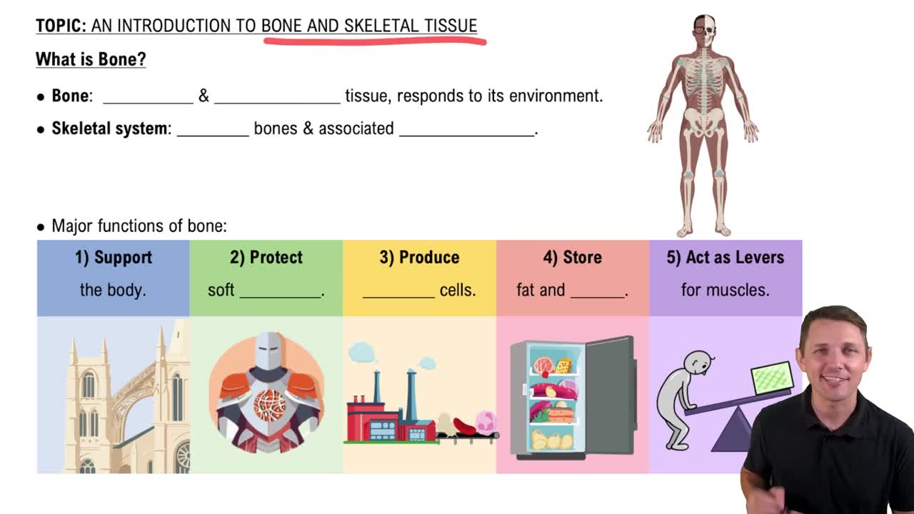
Support, protection, movement/leverage, mineral storage (calcium and phosphate), and blood cell formation in red marrow; yellow marrow stores fat.
Osteocytes are mature bone cells in lacunae;
osteoblasts build bone;
osteoclasts break down bone.
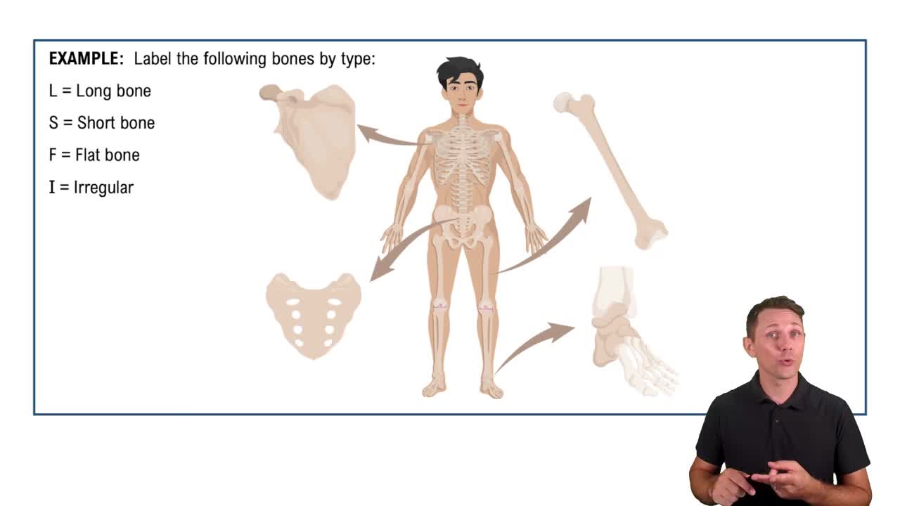
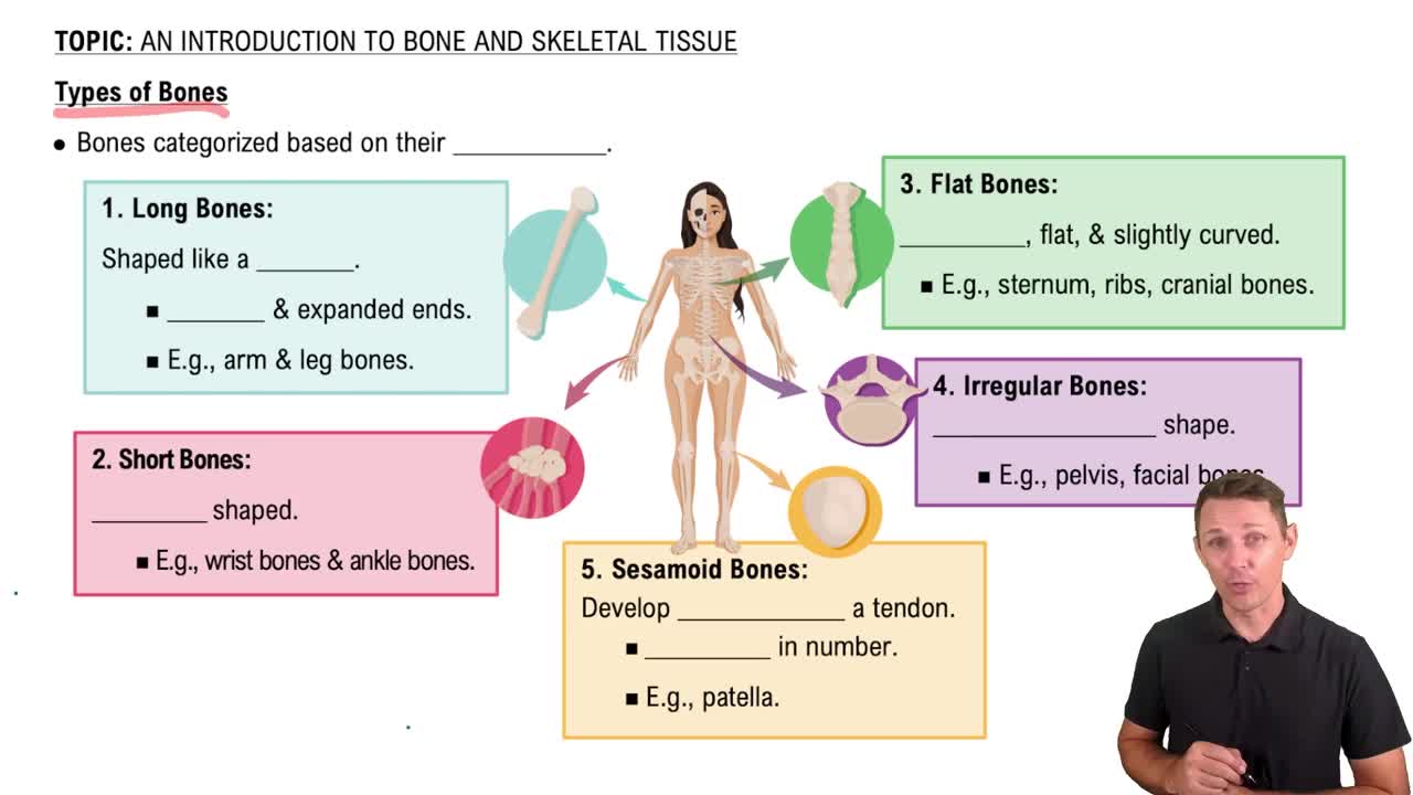
Long (femur), short (carpals), flat (sternum), irregular (vertebra), sesamoid (patella).
Osteons (Haversian systems) are the functional subunits of compact bone.
Central canals run longitudinally through osteons carrying blood vessels and nerves; perforating (Volkmann’s) canals run transversely connecting central canals and periosteum.
Perforating (Sharpey’s) fibers anchor the periosteum to the bone surface.
Circumferential lamellae wrap around bone circumference; concentric lamellae form rings around central canals; interstitial lamellae fill spaces between osteons.
Trabeculae and marrow-filled spaces; it lacks typical osteons.
Canaliculi connect lacunae, allowing communication between osteocytes.
The skull, vertebral column, and thoracic cage form the axial skeleton.
Cervical: 7; Thoracic: 12; Lumbar: 5 vertebrae.
Small bodies, transverse foramina, often bifid spinous processes.
C1 is the atlas; C2 is the axis with the dens (odontoid process) allowing head rotation.
7 pairs of true ribs, 5 pairs of false ribs, ribs 11–12 are floating ribs with no anterior attachment.
Axial skeleton: skull, vertebral column, thoracic cage. Appendicular skeleton: pectoral and pelvic girdles plus limbs.
The glenoid cavity faces laterally; the spine is posterior.
Ulna is medial (pinky side); radius is lateral (thumb side) in anatomical position.
Each hip bone forms from the ilium, ischium, and pubis.
Female pelvis is wider, shallower, with larger pelvic inlet/outlet and wider subpubic angle; male pelvis is narrower and deeper.
The pubic symphysis, a fibrocartilaginous joint.
Structural: fibrous, cartilaginous, synovial. Functional: synarthrosis (immovable), amphiarthrosis (slightly movable), diarthrosis (freely movable).
No, fibrous joints lack a synovial cavity.
Sutures (immovable), syndesmoses (slight movement), gomphoses (tooth in socket, immovable).
Synchondroses use hyaline cartilage (immovable); symphyses use fibrocartilage (slightly movable).
Yes, synovial joints have a fluid-filled cavity and are freely movable.
Flexion/extension (sagittal), abduction/adduction (frontal), rotation (transverse), circumduction (circular).
Plane/gliding (intercarpal), hinge (elbow), pivot (atlantoaxial), condyloid/ellipsoid (knuckles), saddle (thumb carpometacarpal), ball-and-socket (shoulder/hip).
Supraspinatus, infraspinatus, teres minor, and subscapularis.
Epimysium surrounds whole muscle; perimysium surrounds fascicles; endomysium surrounds individual muscle fibers.
A bundle of skeletal muscle fibers.
Branched cells joined by intercalated discs; striated and involuntary.
The plasma membrane of a muscle fiber.
The sarcoplasmic reticulum stores and releases calcium ions.
The sarcomere, from one Z disc to the next.
Motor neuron releases acetylcholine; action potential spreads along sarcolemma and T tubules; sarcoplasmic reticulum releases Ca²⁺; calcium binds troponin, exposing actin sites; myosin binds actin and contracts using ATP; calcium pumped back and muscle relaxes.