 Back
Anatomy & Physiology: Bone Structure and Growth
 3:56

Terms in this set (29)
Thin layer of cartilage covering an epiphysis; reduces friction and acts as a shock absorber.
Where two bone surfaces meet.
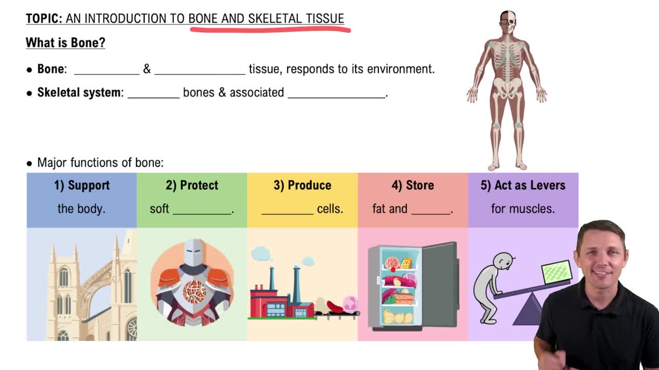
Hard, dense connective tissue that forms the structural elements of the skeleton.
Channels within the bone matrix housing osteocyte cytoplasmic extensions for communication and nutrient exchange.
Semi-rigid connective tissue found on the skeleton where flexibility and smooth surfaces support movement.
Longitudinal channel in the center of each osteon containing blood vessels, nerves, and lymphatic vessels; also called the Haversian canal.
Dense osseous tissue that can withstand compressive forces.
Tubular shaft running between the proximal and distal ends of a long bone.
Layer of spongy bone sandwiched between two layers of compact bone in flat bones.
Process where bone forms by replacing hyaline cartilage.
Delicate membranous lining of a bone’s medullary cavity.
Sheet of hyaline cartilage in the metaphysis of an immature bone; replaced by bone tissue as the bone grows in length.
Wide section at each end of a long bone; filled with spongy bone and red marrow.
Blood clot that forms at the site of a broken bone.
Production of blood cells occurring in the red marrow of bones.
Process by which bone forms directly from mesenchymal tissue.
Spaces in bone that house osteocytes.
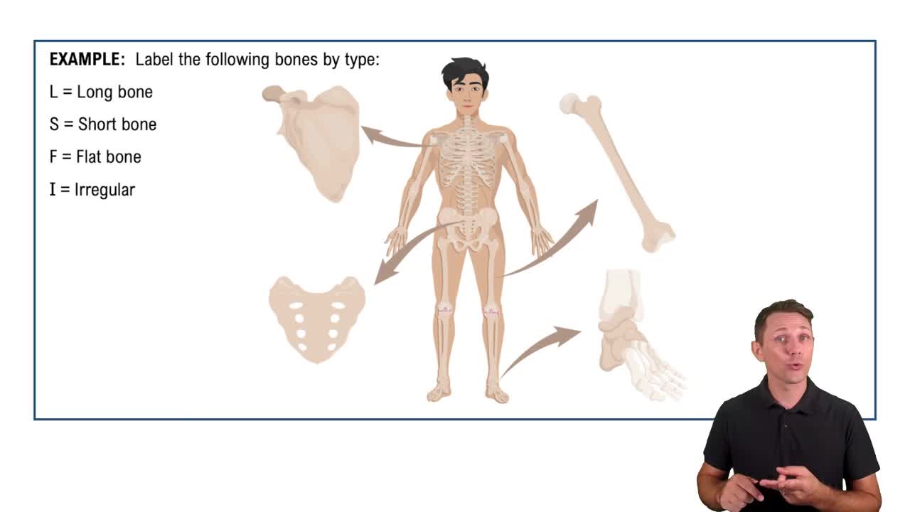
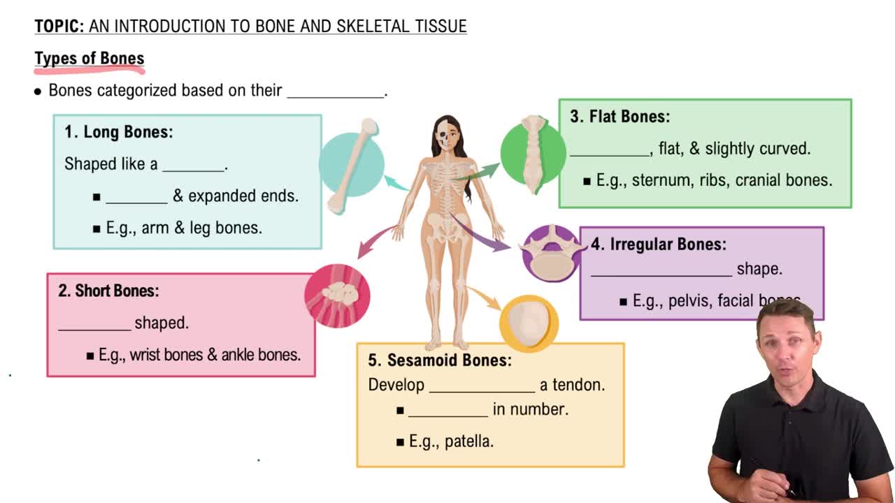
Cylinder-shaped bone longer than it is wide; functions as a lever.
Hollow region of the diaphysis filled with yellow marrow.
Cell responsible for forming new bone.
Cell responsible for resorbing bone.
Primary cell in mature bone responsible for maintaining the matrix.
Undifferentiated bone cell with high mitotic activity; differentiates into osteoblasts.
Basic structural unit of compact bone made of concentric layers of calcified matrix.
Fibrous membrane covering the outer surface of bone and continuous with ligaments.
Region of the epiphyseal plate producing new chondrocytes for longitudinal growth.
Connective tissue in bone interior where hematopoiesis takes place.
Trabeculated osseous tissue supporting shifts in weight distribution.
Connective tissue in bone interior where fat is stored.