 Back
Anatomy & Physiology: Bones, Skeleton, and Joints Study Guide
 3:56

Terms in this set (69)
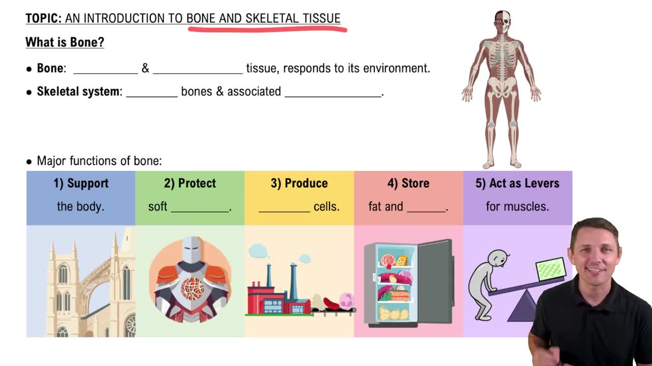
Support, protection, movement, mineral storage, blood cell formation, and energy storage.
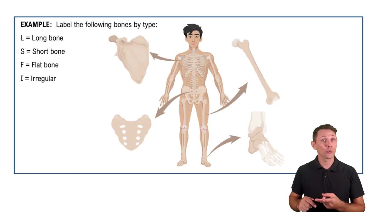
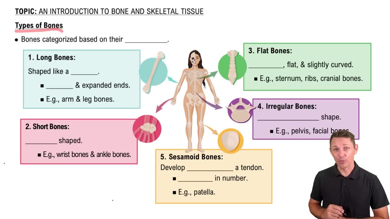
Long bones (longer than wide), short bones (cube-shaped), flat bones (thin, flat), irregular bones (complex shapes), and sesamoid bones (embedded in tendons).
Red bone marrow produces blood cells; found in spongy bone of flat bones and epiphyses of long bones; decreases with age, replaced by yellow marrow.
Yellow marrow stores fat; found in the medullary cavity of long bones; increases with age as red marrow decreases.
Diaphysis (shaft), epiphyses (ends), medullary cavity, articular cartilage, periosteum, and endosteum.
Bone markings serve as sites for muscle attachment, joint formation, and passageways for nerves and blood vessels.
Osteogenic cells: stem cells; osteoblasts: build bone; osteocytes: maintain bone; bone lining cells: regulate calcium; osteoclasts: resorb bone.
It becomes an osteocyte, a mature bone cell embedded in the matrix.
An osteon is the structural unit of compact bone, consisting of concentric lamellae around a central canal containing blood vessels and nerves.
Spongy bone has trabeculae and is lighter, supporting marrow; compact bone is dense, providing strength and protection.
Bone forms directly from mesenchymal tissue; forms flat bones like the skull; involves one ossification center.
Bone forms by replacing hyaline cartilage; forms most bones; involves primary and secondary ossification centers.
Interstitial growth increases bone length; appositional growth increases bone thickness.
Zones of proliferation, hypertrophy, calcification, and ossification allow new cartilage to be replaced by bone, lengthening the bone.
Growth hormone is most important in childhood; others include thyroid hormone and sex hormones; imbalances affect bone length.
Continuous process of bone resorption by osteoclasts and bone deposition by osteoblasts to maintain bone strength and mineral homeostasis.
Resorption: osteoclasts break down bone; deposition: osteoblasts build new bone matrix.
Negative feedback loop for calcium homeostasis and mechanical stress response that strengthens bone where needed.
Parathyroid hormone increases blood calcium by stimulating resorption; calcitonin lowers blood calcium by inhibiting resorption.
Bone mass loss leading to fragility; caused by hormonal changes, age, poor diet, inactivity; risk factors include age, gender, and lifestyle.
Bone density peaks around age 30.
Process of bone tissue formation.
Closes around 18-21 years, becoming the epiphyseal line.
Classified by position, completeness, orientation, and skin penetration.
Hematoma formation, fibrocartilaginous callus formation, bony callus formation, and bone remodeling.
Includes skull, vertebral column, and rib cage; totals 80 bones.
Supports and protects brain, spinal cord, and organs; provides attachment for muscles.
Protects brain, supports sensory organs, and provides attachment for muscles.
Cranium: protects brain, 8 bones; facial bones: form face structure, 14 bones.
Lighten skull, produce mucus, and enhance voice resonance.
Sphenoid bone because it articulates with all other cranial bones.
Does not articulate with any other bone; supports the tongue.
Mandible.
Maxilla because it articulates with all other facial bones except mandible.
Immovable joints between skull bones; mandible is not united by sutures.
Cervical (7), thoracic (12), lumbar (5), sacrum (5 fused), coccyx (4 fused).
Cervical region.
Absorb shock and allow flexibility between vertebrae.
Disc nucleus protrudes through annulus fibrosus due to injury or degeneration.
Infants have a single C-shaped curve; adults have S-shaped curves for balance.
Includes limbs and girdles; totals 126 bones.
Facilitates movement and supports limbs.
Clavicle and scapula.
Humerus, radius, ulna, carpals, metacarpals, phalanges.
Two hip bones (each made of ilium, ischium, pubis).
Femur, patella, tibia, fibula, tarsals, metatarsals, phalanges.
Tibia and talus.
Femur.
Synarthrosis (immovable), amphiarthrosis (slightly movable), diarthrosis (freely movable).
Fibrous, cartilaginous, and synovial joints.
Bones joined by dense fibrous connective tissue; mostly immovable.
Sutures (immovable), syndesmoses (slightly movable), gomphoses (immovable).
Bones joined by cartilage; allow limited movement.
Synchondroses (hyaline cartilage, immovable, e.g. epiphyseal plate), symphyses (fibrocartilage, slightly movable, e.g. intervertebral discs).
Articular cartilage, synovial cavity, synovial fluid, joint capsule, ligaments.
Lubricates and nourishes cartilage; viscous fluid rich in hyaluronic acid.
Fluid-filled sacs that reduce friction between tendons and bones or skin.
Elongated bursae that wrap around tendons to reduce friction.
Articular surface shape, ligament number and location, muscle tone.
Hyaline cartilage covering bone ends; reduces friction and absorbs shock.
Fibrous: mostly synarthrosis; cartilaginous: synarthrosis or amphiarthrosis; synovial: diarthrosis.
Fibrocartilage pads that improve fit and absorb shock in synovial joints.
Gliding, angular, rotation, special movements like supination and opposition.
Plane (wrist), hinge (elbow), pivot (atlas-axis), condylar (knuckle), saddle (thumb), ball-and-socket (shoulder).
Sprain: ligament injury; dislocation: bone displacement from joint.
Joint pain and stiffness due to cartilage wear; caused by aging and joint overuse.
Autoimmune inflammation causing joint pain and deformity.
Uric acid crystal buildup causing joint inflammation and pain.
Bacterial infection from tick bite causing joint pain and inflammation.