Skip to main content
Anatomy & Physiology
My Course
Learn
Exam Prep
AI Tutor
Study Guides
Textbook Solutions
Flashcards
Explore
Try the app
My Course
Learn
Exam Prep
AI Tutor
Study Guides
Textbook Solutions
Flashcards
Explore
Try the app
Back
Anatomy & Physiology: Cardiovascular, Lymphatic, and Immune Systems
You can tap to flip the card.
Pulmonary circuit function
You can tap to flip the card.
👆
Pulmonary circuit function
Right side of heart receives oxygen-poor blood from body tissues and pumps it to lungs to eliminate CO2 and pick up O2.
Track progress
Control buttons has been changed to "navigation" mode.
1/30
Recommended videos
Guided course
3:38
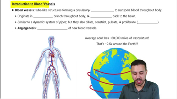
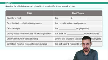
Introduction to Blood Vessels Example 1
6760
views
136
rank
Guided course
4:59
Blood Vessels
9707
views
218
rank
2
comments
Guided course
2:04
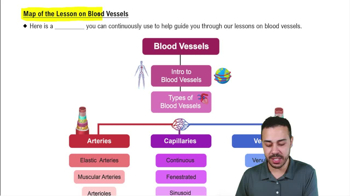
Map of the Lesson on Blood Vessels
6924
views
218
rank
1
comments
Terms in this set (30)
Hide definitions
Pulmonary circuit function
Right side of heart receives oxygen-poor blood from body tissues and pumps it to lungs to eliminate CO2 and pick up O2.
Systemic circuit function
Left side of heart receives oxygenated blood from lungs and pumps it to body tissues to deliver O2 and pick up CO2.
Heart wall layers
Epicardium (visceral serous pericardium), myocardium (cardiac muscle with fibrous skeleton), and endocardium (endothelial lining).
Atrioventricular (AV) valves
Prevent backflow into atria during ventricular contraction; tricuspid valve on right, mitral (bicuspid) valve on left.
Semilunar (SL) valves
Prevent backflow from arteries into ventricles; pulmonary valve on right, aortic valve on left; each has three crescent-shaped cusps.
Coronary circulation
Supplies blood to heart muscle; includes left and right coronary arteries and cardiac veins draining into coronary sinus.
Intrinsic conduction system sequence
SA node → AV node → AV bundle (bundle of His) → right and left bundle branches → Purkinje fibers.
Pacemaker potential
Unstable resting potential in pacemaker cells caused by closing K+ channels and opening slow Na+ channels, leading to gradual depolarization.
Cardiac cycle phases
Includes ventricular filling, isovolumic contraction, ventricular ejection, and isovolumic relaxation.
Frank-Starling law of the heart
Stroke volume increases with increased preload due to greater stretch of cardiac muscle fibers before contraction.
Tunica intima
Innermost blood vessel layer; endothelium and subendothelial layer; continuous with heart endocardium.
Tunica media
Middle vessel layer; mostly circular smooth muscle; controls vasoconstriction and vasodilation.
Tunica externa
Outermost vessel layer; collagen fibers that protect and anchor vessels; contains vasa vasorum in large vessels.
Elastic arteries
Large arteries near heart with thick walls and elastin; act as pressure reservoirs to maintain continuous blood flow.
Capillary types
Continuous (least permeable), fenestrated (pores for filtration), and sinusoidal (most permeable, found in liver, bone marrow).
Precapillary sphincters
Smooth muscle cuffs that regulate blood flow into true capillaries based on local chemical conditions.
Venous valves
Prevent backflow of blood in veins; especially abundant in limbs; assist venous return to heart.
Vascular anastomoses
Interconnections between blood vessels providing alternate pathways for blood flow to ensure tissue perfusion.
Net filtration pressure (NFP)
Determines fluid movement across capillaries; positive at arterial end (filtration), negative at venous end (reabsorption).
Lymphatic system functions
Returns excess interstitial fluid to blood, transports fats from intestines, and filters lymph through lymph nodes.
Lymphatic capillaries
Blind-ended vessels with one-way minivalves; highly permeable to proteins, debris, pathogens, and cancer cells.
Phagocytosis process
Phagocyte binds particle, encloses it in phagosome, fuses with lysosome to form phagolysosome, then digests contents.
Natural killer (NK) cells
Nonphagocytic lymphocytes that kill virus-infected and cancer cells by inducing apoptosis and secreting inflammatory chemicals.
Inflammation benefits
Prevents pathogen spread, disposes of debris, alerts immune system, and sets stage for tissue repair.
Complement system
Plasma proteins activated via classical, lectin, or alternative pathways; promote inflammation, opsonization, and cell lysis.
Fever advantages
Enhances immune cell migration, increases metabolic rate and repair, and inhibits bacterial growth by reducing metal ion availability.
MHC proteins
Self-antigens on cell surfaces; Class I present endogenous antigens to CD8 T cells; Class II present exogenous antigens to CD4 T cells.
Helper T cells functions
Activate CD8 cytotoxic T cells, amplify innate defenses, and mobilize other immune cells via cytokine secretion.
Cytotoxic T cells
Directly kill infected, cancerous, or foreign cells by releasing perforins and granzymes to induce apoptosis.
Active vs passive humoral immunity
Active: B cells produce antibodies after antigen exposure; Passive: antibodies received from another source, no memory formed.
 BackBack
 3:38