 Back
Anatomy & Physiology: Cardiovascular System Exam Prep
 6:13

Terms in this set (30)
Stroke volume is the amount of blood ejected by the left ventricle per beat. Cardiac output is the volume of blood the heart pumps per minute, calculated as stroke volume multiplied by heart rate.
The three factors are preload (ventricular filling), contractility (strength of contraction), and afterload (resistance to ejection). Increasing preload or contractility increases stroke volume; increasing afterload decreases it.
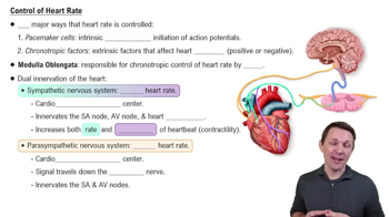
Beta blockers block beta-1 adrenergic receptors, reducing heart rate and contractility. This lowers cardiac output and oxygen demand. Generic examples include propranolol. They help performance anxiety by reducing sympathetic effects.
CHF is a condition where the heart cannot pump blood effectively, leading to fluid buildup. It can be left-sided (pulmonary congestion) or right-sided (systemic venous congestion).
Left-sided failure causes pulmonary edema and dyspnea due to backup in lungs. Right-sided failure causes peripheral edema and jugular venous distension from systemic venous backup.
Left-sided causes include ischemic heart disease and hypertension. Right-sided failure often results from left-sided failure or pulmonary diseases like COPD.
Treatments include diuretics (reduce fluid overload), ACE inhibitors (reduce afterload), beta blockers (reduce heart rate), and digitalis (increase contractility). Therapy varies by dominant side.
MI diagnosis uses ECG changes (ST elevation, Q waves) and biomarkers like troponin. Non-ischemic pain lacks these specific changes.
Atrial repolarization is obscured because it occurs during ventricular depolarization, which produces a larger QRS complex that masks it.
Starts with endothelial injury, followed by lipid accumulation, inflammation, foam cell formation, and fibrous cap development. Plaque stability affects risk of rupture and thrombosis.
Cardiac tamponade is fluid accumulation in the pericardium that compresses the heart, impairing filling and reducing cardiac output.
Signs include hypotension, jugular venous distension, and muffled heart sounds (Beck's triad).
To remove fluid and relieve pressure, restoring cardiac filling and output to prevent cardiovascular collapse.
Pericarditis is inflammation of the pericardium, causing chest pain and a pericardial friction rub. ECG may show diffuse ST elevation.
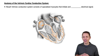
They arise from abnormal automaticity or triggered activity, causing premature beats or arrhythmias when normal pacemaker control is lost.
Caused by atrial remodeling or triggers; leads to irregular ventricular response and reduced cardiac efficiency, increasing stroke risk.
Rate control manages ventricular rate without restoring sinus rhythm; rhythm control aims to restore and maintain normal rhythm.
Atrial flutter shows regular sawtooth waves from a reentrant circuit; atrial fibrillation shows irregular, chaotic atrial activity.
It causes uncoordinated ventricular contractions, stopping effective cardiac output and leading to loss of consciousness.
Immediate defibrillation followed by CPR restores coordinated contractions and cardiac output.
Risk factors include valve disease and IV drug use. Vegetations form from bacterial colonization and platelet-fibrin deposits on valves.
Antibiotics target causative organisms, given for prolonged duration to eradicate bacteria in vegetations.
Stenosis narrows valve opening, increasing pressure load. Regurgitation causes backward flow, increasing volume load.
Stenosis causes concentric hypertrophy; regurgitation causes eccentric hypertrophy due to volume overload.
Mitral stenosis causes a diastolic murmur and symptoms like dyspnea from pulmonary congestion.
Mitral regurgitation causes a holosystolic murmur and symptoms of fatigue and pulmonary edema.
Aortic stenosis causes a systolic ejection murmur and symptoms like syncope and angina.
Aortic regurgitation causes a diastolic murmur and symptoms of dyspnea and fatigue.
Tricuspid stenosis causes a diastolic murmur and symptoms of systemic venous congestion.
Tricuspid regurgitation causes a holosystolic murmur and signs of right heart failure.