 Back
Anatomy & Physiology Chapter 5: Integumentary System Study Guide
 06:54

Terms in this set (65)
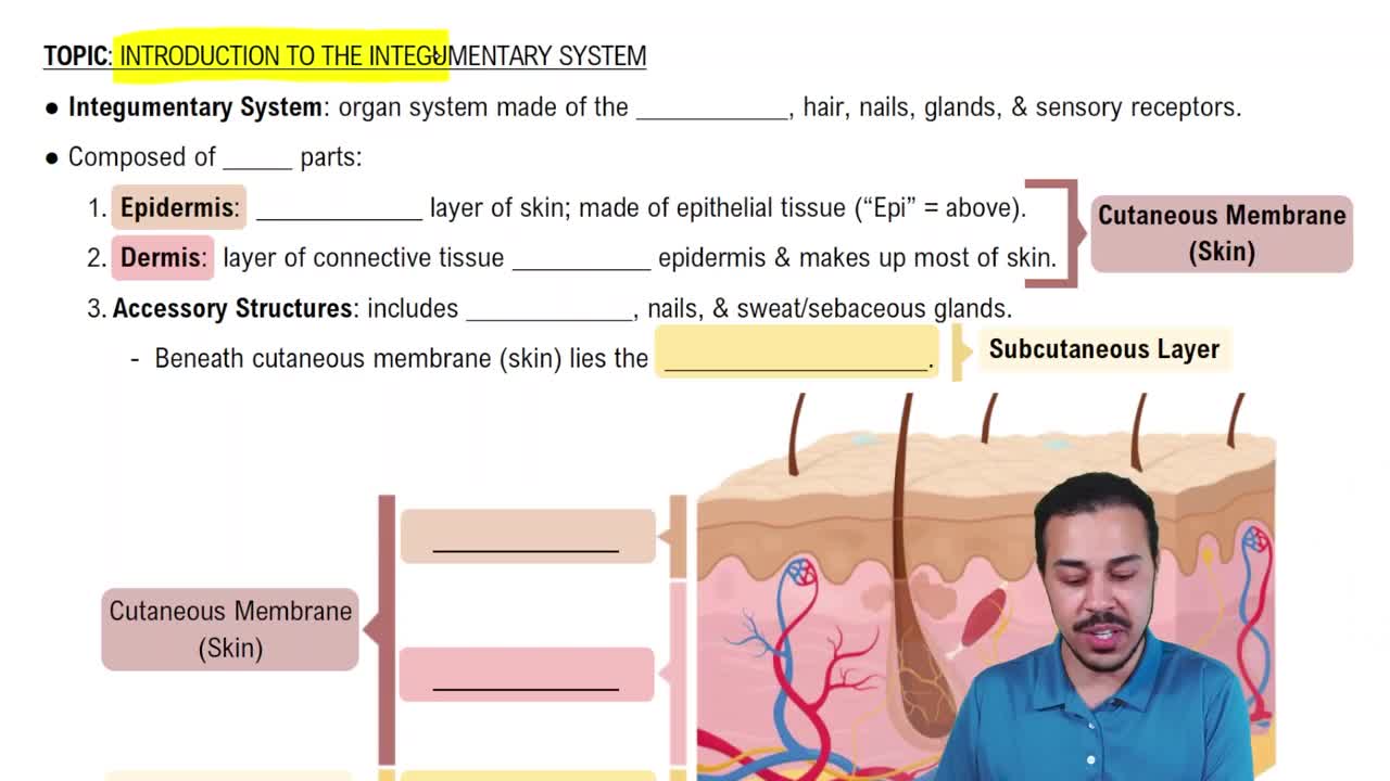
The epidermis and the dermis.
Located beneath the dermis; composed mainly of areolar and adipose connective tissue.
- Stores fat
- Provides insulation
- Anchors skin to underlying structures
Keratinized stratified squamous epithelium.
Produce keratin, a protein that strengthens skin and makes it waterproof.
Produce melanin, the pigment that protects against UV radiation.
Act as immune cells that detect pathogens and activate immune responses.
Serve as touch receptors by associating with sensory nerve endings.
Keratinocytes are dominant; they produce keratin.
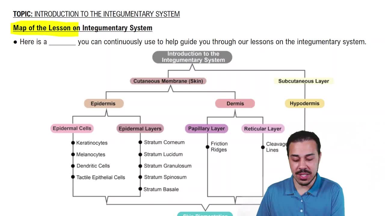
Stratum basale, stratum spinosum, stratum granulosum, stratum lucidum (only in thick skin), stratum corneum.
Stratum lucidum.
Stratum basale.
Stratum basale.
Stratum lucidum (thick skin) and stratum corneum.
Stratum granulosum.
Stratum spinosum.
Palms of hands and soles of feet.
- Thick skin has all five epidermal layers and no hair follicles or sebaceous glands.
- Thin skin lacks stratum lucidum and contains hair follicles, sebaceous, and sweat glands.
Papillary layer: areolar connective tissue; reticular layer: dense irregular connective tissue.
Reticular layer, making up about 80% of the dermis.
Fingerlike projections of the papillary dermis that increase surface area for nutrient exchange and strengthen attachment to epidermis.
Dermal papillae form ridges on the skin surface, creating unique fingerprint patterns.
Melanin.
Melanocytes in the stratum basale.
Protects skin cells from UV radiation damage.
Hemoglobin in red blood cells.
Carotene.
- Hair
- Nails
- Sweat glands
- Sebaceous glands
Hair and nails.
Contracts to cause hair to stand up, producing 'goosebumps' and helping trap heat.
Medulla, cortex, cuticle.
Internal root sheath, external root sheath, connective tissue sheath.
- Protection
- Sensation
- Heat retention
- Nail plate: visible hard part
- Nail bed: skin beneath plate
- Nail matrix: growth region
- Nail folds: skin around nail
- Eponychium (cuticle): skin at nail base
- Hyponychium: skin under free edge
Nail matrix.
Located all over body; secrete watery sweat; function in thermoregulation.
Located in axillary and genital areas; secrete milky sweat; function in scent release.
Associated with hair follicles; secrete oily sebum; lubricate skin and hair.
Eccrine sweat glands.
Apocrine sweat glands and sebaceous glands.
No, because thick skin lacks hair follicles which sebaceous glands associate with.
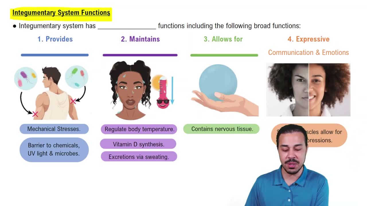
- Protection
- Temperature regulation
- Sensation
- Vitamin D synthesis
- Acid mantle (low pH)
- Defensins (antimicrobial peptides)
Stratum corneum.
'Bricks' are keratinized cells; 'mortar' is the lipid matrix preventing water loss.
- Dendritic cells in epidermis
- Macrophages in dermis
- Vasodilation of dermal blood vessels
- Activation of sweat glands
Vitamin D.
Exposure to UVB radiation from sunlight.
Needed for calcium absorption and bone health.
Basal cells in the stratum basale.
Keratinocytes in the stratum spinosum.
Melanocytes in the stratum basale.
Basal cell carcinoma.
Melanoma.
Estimates the percentage of body surface area affected by burns for treatment decisions.
Only the epidermis.
Epidermis and part of the dermis.
Entire thickness of the skin including epidermis, dermis, and often subcutaneous tissue; also called full-thickness burns.
Contains all five epidermal layers, lacks hair follicles and sebaceous glands.
Melanocytes.
Protects skin cells from UV damage by absorbing radiation.
Eccrine sweat glands.
Cells are dead and lack nuclei, so they cannot divide or become cancerous.
Weakens epidermal protection and causes structural defects in hair and nails.