 Back
Anatomy & Physiology Exam 1
 05:04

Terms in this set (21)
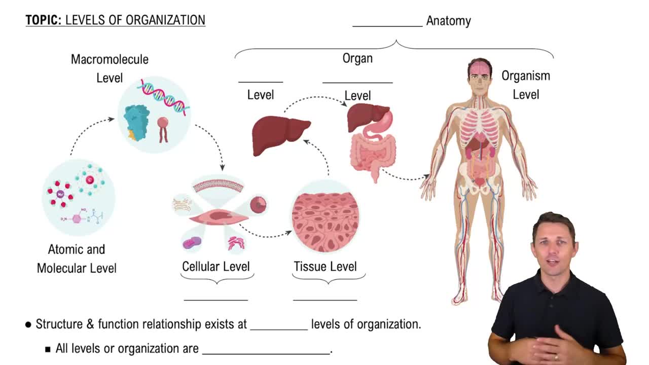
Atoms → molecules → cells → tissues → organs → organ systems → organism.
Anatomy is the study of structure (form), while physiology is the study of function.
Standing, facing forward, arms at sides, palms forward, feet slightly apart; a shared reference frame for directional terms.
Proximal: closer to the trunk or point of origin; Distal: farther from the trunk or point of origin, used for limbs and tubular organs.
Sagittal: divides body into left and right; Midsagittal: equal left and right halves; Parasagittal: unequal left and right sections.
Dorsal cavity: cranial (brain) + vertebral (spinal cord); Ventral cavity: thoracic (lungs, heart) + abdominopelvic (digestive, urinary, reproductive organs).
Parietal layer lines the cavity wall; Visceral layer covers the organ surface; separated by serous fluid.
Moves 3 Na⁺ ions out and 2 K⁺ ions into the cell per ATP, maintaining ion gradients and membrane potential.
Passive transport requires no energy and moves substances down their gradient; Active transport requires ATP and moves substances against their gradient.
Hypotonic: water enters cell, causing swelling; Isotonic: no net water movement; Hypertonic: water leaves cell, causing shrinkage.
Transcription: DNA → mRNA in nucleus; Translation: mRNA + tRNA → polypeptide at ribosomes.
High cellularity, free apical surface, basal surface attached to basement membrane; classified by layers and cell shape.
Single layer of flat cells; allows diffusion of gases; found in alveoli, capillaries, and serous membranes.
Dense regular: collagen fibers parallel, resists tension in one direction (tendons, ligaments); Dense irregular: collagen fibers in multiple directions, resists tension in many directions (dermis, organ capsules).
Fibroblasts produce collagen and ground substance; Macrophages phagocytose debris and bacteria.
Skeletal: voluntary, striated; Cardiac: involuntary, striated, intercalated discs; Smooth: involuntary, nonstriated, found in uterus and organs.
Tight junctions: seal spaces between cells; Desmosomes: mechanical strength; Gap junctions: allow ion/small molecule flow for electrical coupling.
Negative feedback opposes change to restore set point (e.g., sweating); Positive feedback amplifies change until an endpoint (e.g., childbirth).
Actin filaments: thin, interact with myosin for movement; Microtubules: transport highways, form cilia/flagella; Intermediate filaments: mechanical strength, nuclear lamina.
Produce ATP; abundant in cells with high energy demand like muscle cells.
Phagocytosis: cell eating of large particles/bacteria; Pinocytosis: cell drinking of extracellular fluid droplets.