 Back
Anatomy & Physiology Exam 1 Study Guide
 2:36

Terms in this set (37)
Transport of gases, nutrients, and wastes; regulation of pH and temperature; protection via clotting and immune responses.
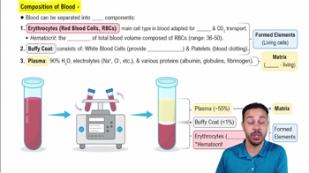
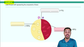
Plasma (~55%), buffy coat (leukocytes and platelets, <1%), and erythrocytes (~45%).
Cells and cell fragments: erythrocytes (red blood cells), leukocytes (white blood cells), and platelets.
The percentage of blood volume composed of erythrocytes.
Blood type is determined by specific antigens on erythrocytes; antibodies in plasma react against foreign antigens.
A blood group antigen; presence (+) or absence (−) affects blood compatibility and pregnancy risks.
Hemoglobin is a protein with four heme groups containing iron; it carries oxygen in erythrocytes.
Erythrocytes are red blood cells; erythropoiesis is their production in the red bone marrow.
Leukocytes are white blood cells; leukopoiesis is their production in the red bone marrow.
Granulocytes: neutrophils, eosinophils, basophils. Agranulocytes: lymphocytes and monocytes.
Neutrophils: phagocytosis; Lymphocytes: B cells produce antibodies, T cells attack infected cells; Monocytes: become macrophages; Eosinophils: combat parasites; Basophils: release histamine.
Platelets are cell fragments formed from megakaryocytes in bone marrow; they aid in clotting.
Vascular spasm, platelet plug formation, and coagulation (clotting) to stop bleeding.
Endocrine: hormones into blood; Exocrine: secretions via ducts; Paracrine: local cell signaling; Autocrine: self-signaling.
Hormones travel through the bloodstream to target cells.
Receptors on target cells bind hormones to trigger specific cellular responses.
Steroid hormones are lipid soluble, derived from cholesterol, act inside cells on DNA; protein/amino acid hormones are water soluble, act on cell surface receptors.
Water soluble hormone binds receptor → activates G protein → activates adenylate cyclase → produces cAMP → activates protein kinases → cellular response.
Up regulation: increase in receptors due to low hormone levels; Down regulation: decrease in receptors due to high hormone levels.
Hormones with opposing effects, e.g., insulin lowers blood glucose, glucagon raises it.
Posterior pituitary: stores/releases oxytocin and ADH; Anterior pituitary: produces hormones like GH, ACTH, TSH, FSH, LH, and prolactin.
High blood glucose triggers insulin release, which lowers glucose, reducing insulin secretion.
Oxytocin release during childbirth intensifies contractions, causing more oxytocin release.
Fibrous pericardium: outer protective layer; Serous pericardium: parietal and visceral layers with fluid that reduces friction.
Epicardium: outer layer; Myocardium: muscular middle layer; Endocardium: inner lining.
Atria: receive blood; Ventricles: pump blood out of the heart.
Pulmonary circuit: right heart to lungs and back; Systemic circuit: left heart to body and back.
Prevent backflow of blood and ensure one-way flow through the heart.
P wave: atrial depolarization; QRS complex: ventricular depolarization; T wave: ventricular repolarization.
Body → vena cava → right atrium → right ventricle → pulmonary artery → lungs → pulmonary veins → left atrium → left ventricle → aorta → body.
Blood supply to the heart muscle via coronary arteries and veins.
Sinoatrial (SA) node: primary pacemaker; Atrioventricular (AV) node: delays impulse before ventricles contract.
Arteries carry blood away from the heart; veins carry blood toward the heart.
The central compartment of the thoracic cavity containing the heart, great vessels, trachea, and esophagus.
Includes SA node, AV node, AV bundle, bundle branches, and Purkinje fibers that coordinate heartbeats.
Preload is the degree of ventricular stretch before contraction; increased preload increases stroke volume.
Sympathetic: increases heart rate; Parasympathetic: decreases heart rate.