 Back
Anatomy & Physiology Exam 2 Study Guide - Bone, Joints, and Nervous System
 3:56

Terms in this set (63)
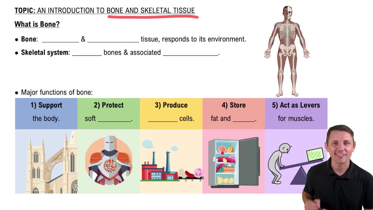
The skeletal system provides support, protection of organs, movement via muscle attachment, mineral storage (calcium and phosphate), blood cell production in marrow, and energy storage in yellow marrow.
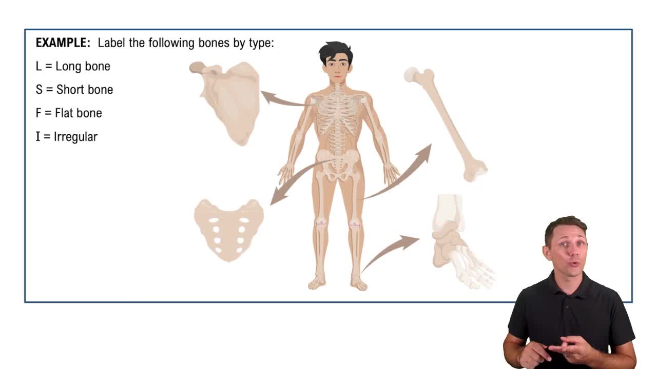
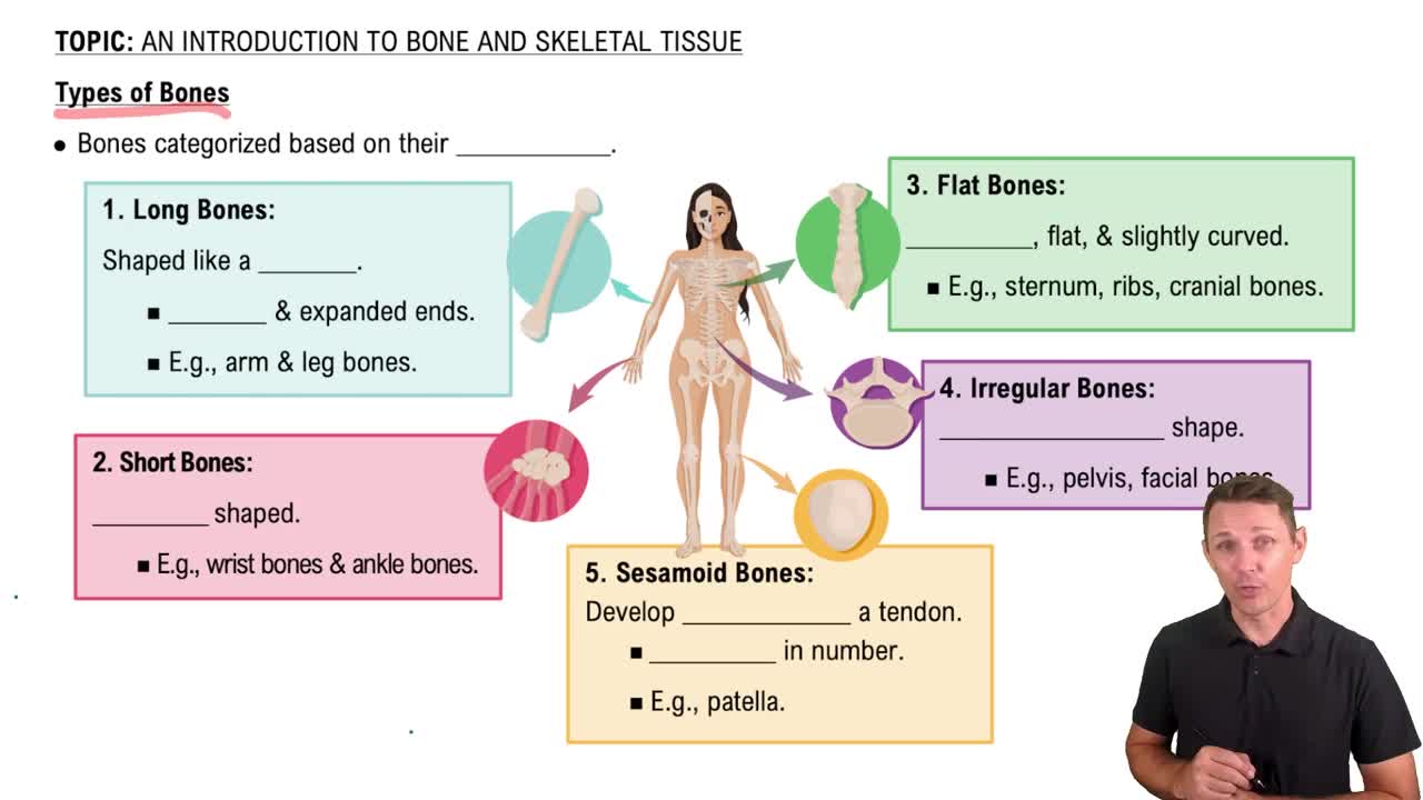
Long bones (e.g., femur), short bones (e.g., carpals), flat bones (e.g., sternum), and irregular bones (e.g., vertebrae).
Osteoblasts build bone, osteocytes maintain bone matrix, and osteoclasts resorb (break down) bone.
Consists of diaphysis (shaft), epiphyses (ends), medullary cavity (marrow), compact bone, spongy bone, periosteum, and endosteum.
Two matrices: organic matrix (collagen fibers for flexibility) and inorganic matrix (hydroxyapatite crystals for hardness and strength).
Leads to brittle bones because collagen provides flexibility; without it, bones become fragile and prone to fractures (osteogenesis imperfecta).
Osteoblasts synthesize and deposit new bone matrix, while osteoclasts break down bone tissue to release minerals.
An osteon includes central canal (blood vessels), lamellae (concentric rings), lacunae (osteocyte spaces), and canaliculi (channels for nutrient exchange).
Growth in length occurs by chondrocyte proliferation, hypertrophy, calcification, and ossification at the epiphyseal plate.
Mesenchymal cells differentiate into osteoblasts, secrete bone matrix, form spicules, and develop into flat bones like the skull.
Osteoblasts produce and mineralize bone matrix directly from mesenchymal tissue without a cartilage precursor.
Cartilage model forms, chondrocytes hypertrophy, matrix calcifies, blood vessels invade, osteoblasts replace cartilage with bone.
Intramembranous ossification forms bone directly from mesenchyme; endochondral ossification uses a cartilage template.
The epiphyseal plate is a cartilage growth plate in children; the epiphyseal line is the ossified remnant in adults after growth ends.
Increased testosterone and estrogen accelerate epiphyseal plate closure, affecting final adult height differences between sexes.
Bone growth in thickness by osteoblasts adding new bone matrix on the surface and osteoclasts resorbing bone inside the medullary cavity.
Include growth hormone, thyroid hormone, sex hormones (testosterone, estrogen), and calcitonin.
Continuous process of bone resorption and deposition to maintain strength, repair microdamage, and regulate calcium levels.
Exercise increases bone deposition by stimulating osteoblast activity, leading to stronger bones compared to sedentary individuals.
Parathyroid hormone increases blood calcium by stimulating osteoclasts; calcitriol enhances calcium absorption in the intestines.
Vitamin C deficiency impairs collagen synthesis weakening bone matrix; vitamin D deficiency reduces calcium absorption causing weak, soft bones.
Hematoma formation, fibrocartilaginous callus formation, bony callus formation, and bone remodeling.
Damage can halt longitudinal growth, potentially causing shorter limb length or deformity.
Ossification is bone tissue formation; calcification is deposition of calcium salts in tissue, which can occur without ossification.
Bone resorption continues but deposition decreases, leading to net bone loss and weaker bones.
Axial skeleton (skull, vertebrae, ribs) and appendicular skeleton (limbs and girdles).
Consists of the ilium, ischium, and pubis fused to form the hip bone.
Includes the clavicle and scapula.
Facial bones form the face (e.g., maxilla, mandible); cranial bones enclose the brain (e.g., frontal, parietal).
Synarthrosis (immovable), amphiarthrosis (slightly movable), and diarthrosis (freely movable).
Connected by dense connective tissue, mostly immovable or slightly movable, e.g., sutures in the skull.
Sutures (skull), syndesmoses (interosseous membrane), and gomphoses (tooth sockets).
Connected by cartilage and allow limited movement.
Synchondroses (hyaline cartilage, e.g., epiphyseal plate) and symphyses (fibrocartilage, e.g., pubic symphysis).
Synovial joints have a fluid-filled cavity allowing free movement; fibrous and cartilaginous joints lack this cavity.
Includes articular cartilage, synovial membrane, joint cavity, ligaments, and fibrous capsule.
Lubricates joint, nourishes cartilage, and absorbs shock.
Include gliding, angular (flexion, extension), rotation, and special movements like supination.
Hinge joints are more stable but less mobile (e.g., elbow); ball-and-socket joints are less stable but more mobile (e.g., shoulder).
Elbow has stronger ligament support and bony congruence; knee has more complex structure and is more prone to injury.
Anterior cruciate ligament (ACL), posterior cruciate ligament (PCL), medial collateral ligament (MCL), lateral collateral ligament (LCL), plus medial and lateral menisci.
Both are ball-and-socket joints; shoulder has greater range of motion but less stability than hip.
Supraspinatus, infraspinatus, teres minor, and subscapularis tendons stabilize the shoulder joint.
Leads to pain, reduced mobility, and increased risk of arthritis due to loss of smooth joint surface.
Bursa cushions joints; tendon sheath surrounds tendons to reduce friction.
High mobility with less stability and shallow glenoid cavity make it prone to dislocation and injury.
Osteoarthritis (wear and tear), rheumatoid arthritis (autoimmune), and gouty arthritis (uric acid crystals).
Divided into central nervous system (CNS) and peripheral nervous system (PNS).
Somatic (voluntary control) and autonomic (involuntary control) nervous systems.
Multipolar, bipolar, and unipolar neurons based on number of processes.
Sensory (afferent), motor (efferent), and interneurons.
CNS: astrocytes, oligodendrocytes, microglia, ependymal cells; PNS: Schwann cells, satellite cells.
Insulating layer around axons that increases action potential speed; formed by oligodendrocytes in CNS and Schwann cells in PNS.
High K+ inside, high Na+ outside; this gradient maintains resting membrane potential.
Leak channels are always open; gated channels open or close in response to stimuli.
Depolarization to threshold, opening of Na+ channels, repolarization by K+ efflux, and hyperpolarization before returning to rest.
Time during which a neuron cannot fire another action potential due to inactivated Na+ channels.
Ca2+ influx triggers neurotransmitter release into synaptic cleft.
Electrical synapses allow direct ion flow; chemical synapses use neurotransmitters for signal transmission.
EPSP depolarizes membrane promoting action potential; IPSP hyperpolarizes membrane inhibiting action potential.
Saltatory conduction jumps between nodes of Ranvier in myelinated axons; continuous conduction moves stepwise along unmyelinated axons.
Myelin sheath destruction slows or blocks action potentials, causing neurological deficits.
Glutamate binding typically causes excitatory postsynaptic potentials, increasing likelihood of action potential.