 Back
BackAnatomy & Physiology Exam 4 Study Guide
 05:17
05:17
Terms in this set (27)
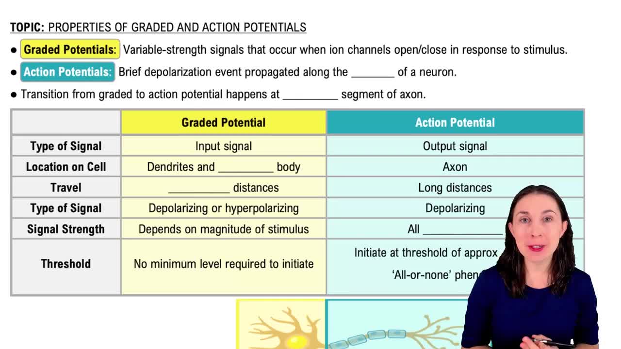
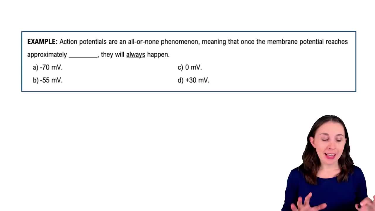
Graded potentials vary in magnitude, are localized, and decrease in strength as they spread. They differ from action potentials which are all-or-none and propagate without decrement.
Graded potentials summate to reach threshold, triggering action potentials that propagate signals over long distances.
Sensory receptors are matched to stimuli such as mechanoreceptors (touch), thermoreceptors (temperature), photoreceptors (light), chemoreceptors (chemicals), and nociceptors (pain).
Action potentials code stimulus intensity by frequency (more frequent firing for stronger stimuli) and by recruiting more neurons for larger stimuli.
CNS includes the brain and spinal cord; PNS includes all nerves outside the CNS, including spinal and cranial nerves.
Dorsal roots carry sensory (afferent) information to the CNS; ventral roots carry motor (efferent) information away from the CNS.
Sensory and motor axons mix after the dorsal and ventral roots merge to form the spinal nerve proper.
Spinal nerves branch into dorsal and ventral rami; the ventral ramus connects with limbs and anterior body wall muscles.
Nerve plexuses allow complex innervation of limb muscles by mixing fibers from multiple spinal nerves, providing redundancy and coordinated control.
Radial nerve controls wrist and finger extension; ulnar nerve controls finger flexion and intrinsic hand muscles.
Femoral nerve controls thigh flexion and leg extension; obturator nerve controls thigh adduction; sciatic nerve controls leg flexion and foot movements.
The neuromuscular junction is the synapse between a motor neuron and muscle fiber; the end plate potential is the depolarization of the muscle membrane that triggers contraction.
Efferent nerves synapse with smooth muscle and glands via varicosities releasing neurotransmitters diffusely rather than at a single junction.
Intrinsic reflexes are innate, automatic responses; learned reflexes are acquired through practice.
Yes, intrinsic reflexes can be modulated by higher brain centers and experience.
Monosynaptic reflexes involve one synapse between sensory and motor neurons; polysynaptic reflexes involve one or more interneurons.
Extrafusal fibers generate force for muscle contraction; intrafusal fibers are part of muscle spindles that detect stretch.
Muscle spindles send action potentials to the CNS about muscle length and rate of stretch, aiding proprioception.
The muscle stretch reflex causes contraction of stretched muscle; reciprocal inhibition relaxes antagonist muscles to allow smooth movement.
The three primary brain vesicles are prosencephalon (forebrain), mesencephalon (midbrain), and rhombencephalon (hindbrain).
Prosencephalon divides into telencephalon and diencephalon; mesencephalon remains undivided; rhombencephalon divides into metencephalon and myelencephalon.
Cerebral hemispheres develop from telencephalon; thalamus from diencephalon; midbrain from mesencephalon; cerebellum from metencephalon; medulla oblongata from myelencephalon.
The diencephalon's walls are formed by the thalamus, the floor by the hypothalamus, the roof by the epithalamus, and the center contains the third ventricle.
CSF cushions the brain, removes waste, and provides nutrients. It is found in ventricles and subarachnoid space and secreted by the choroid plexus.
The meninges are dura mater, arachnoid mater, and pia mater. The arachnoid mater encloses the subarachnoid space where CSF circulates.
Medulla controls autonomic functions; cerebellum coordinates movement and balance; hypothalamus regulates homeostasis and endocrine functions.
Precentral gyrus is the primary motor cortex; postcentral gyrus is the primary somatosensory cortex.