 Back
Anatomy & Physiology: Integumentary and Skeletal System Study Guide
 06:54

Terms in this set (29)
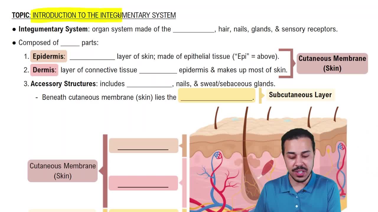
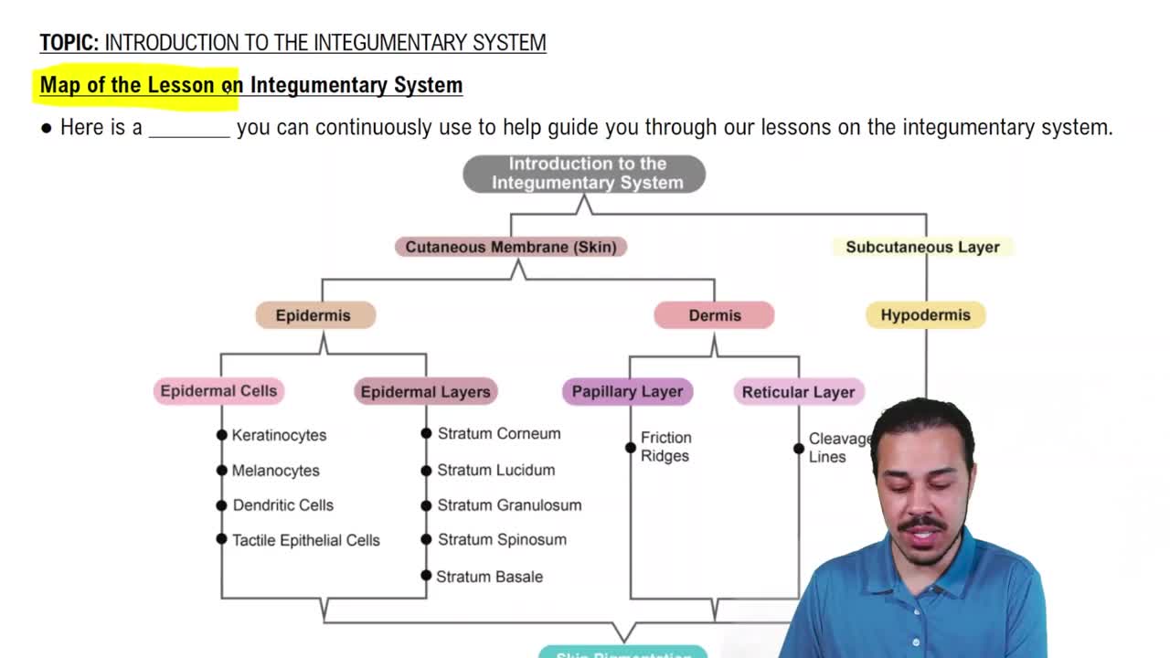
Skin (epidermis and dermis), glands (sweat and oil), hairs, nails, and subcutaneous tissue (adipose tissue beneath skin).
Cutaneous refers to skin layers (epidermis and dermis). Subcutaneous is below skin (hypodermis), not part of skin but anchors it and provides insulation.
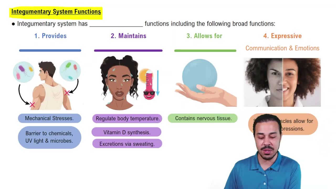
Protection (chemical, physical, biological), temperature regulation, cutaneous sensation, metabolic functions (vitamin D synthesis), blood reservoir, and excretion.
Keratinized stratified squamous epithelial tissue.
Stratum basale: mitotic keratinocytes.
Stratum spinosum: melanocytes, tactile and dendritic cells.
Stratum granulosum: keratin granules, cells dying.
Stratum lucidum: only in thick skin, dead cells.
Stratum corneum: dead keratinocytes, outermost layer.
Keratinocytes: produce keratin.
Melanocytes: produce melanin for UV protection.
Dendritic cells: immune phagocytes.
Tactile cells: sensory receptors.
Thick skin is on palms, soles, and upper back for protection; contains stratum lucidum.
Thin skin lacks stratum lucidum and covers most of the body.
Loose connective tissue (papillary layer) and dense irregular connective tissue (reticular layer).
Papillary layer: blood diffusion, capillaries, nerve endings, touch receptors.
Reticular layer: glands, sensory endings, blood vessels, hair follicles.
Melanin: pigment made in skin.
Hemoglobin: pinkish hue from blood.
Carotene: yellow/orange pigment from diet.
Erythema: redness.
Jaundice: yellowing, liver failure.
Pallor: paleness from low blood flow.
Cyanosis: blue from low oxygen.
Hematoma: bruise.
Hyperpigmentation: brown/black patches.
Eccrine glands: sweat for temperature regulation.
Apocrine glands: puberty onset, foul sweat, axillary and anogenital.
Sebaceous glands: oil secretion, lubrication, acne.
Thermoregulation via sweat, chemical protection by defensin and low pH, biological protection by dendritic cells, and physical barrier by keratinized cells.
Hyaline: support and flexibility, most abundant.
Elastic: collagen and elastic fibers, external ear.
Fibrocartilage: tensile strength, menisci and intervertebral discs.
Support, protection, anchorage for muscles, mineral storage, blood cell production, fat storage, and hormone production (osteocalcin).
Long: femur, humerus.
Short: carpals, tarsals.
Flat: ribs, skull.
Irregular: vertebrae, pelvis.
Red marrow: blood cell production, found in long and flat bones.
Yellow marrow: fat storage, can convert back to red marrow.
Central canal (arteries, veins, nerves), perforating canals, lacunae (osteocyte houses), lamellae (bone layers), and canaliculi (connections).
Osteoblasts: build bone.
Osteocytes: maintain bone.
Osteoclasts: resorb bone.
Intramembranous: from fibrous membrane, forms flat bones.
Endochondral: from hyaline cartilage, forms long bones.
Resting zone, proliferation zone (mitosis), hypertrophic zone (cell enlargement), calcification zone (matrix calcifies), ossification zone (new bone forms).
Appositional: thickness growth by osteoblasts under periosteum.
Interstitial: length growth at epiphyseal plate.
Hematoma formation, soft callus formation, hard callus formation, and bone remodeling.
Nondisplaced vs displaced, complete vs incomplete, compound vs simple, spiral, epiphyseal, depressed, greenstick, comminuted, compression.
Released when blood calcium is low; stimulates osteoclasts to resorb bone and release calcium into blood.
Produced by thyroid parafollicular cells when calcium is high; lowers blood calcium temporarily by inhibiting osteoclasts.
Cell in the bone that handles growth
Osteoblast
Cell in the bone that handles maintenance
Osteocyte
Cell in the bone that handles resorption
Osteoclast