 Back
Anatomy & Physiology: Integumentary System Study Guide
 06:54

Terms in this set (31)
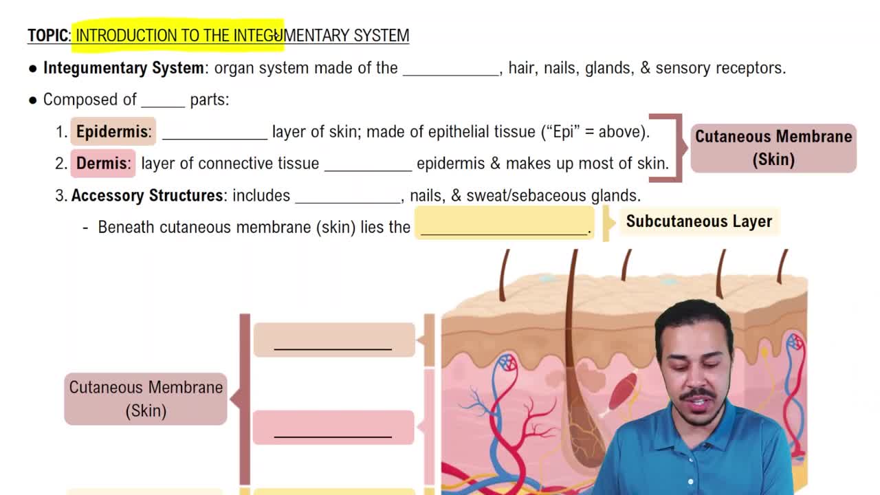
Epidermis: keratinized stratified squamous epithelium.
Dermis: loose and dense connective tissue.
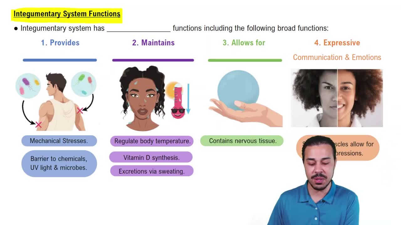
Touch, temperature, pain, vibration, and pressure.
Oxygen and nutrients diffuse from capillaries in the dermis.
Thin skin lacks the stratum lucidum and has 4 epidermal layers; found over most of the body.
Thick skin contains the stratum lucidum and has 5 layers; found on palms and soles.
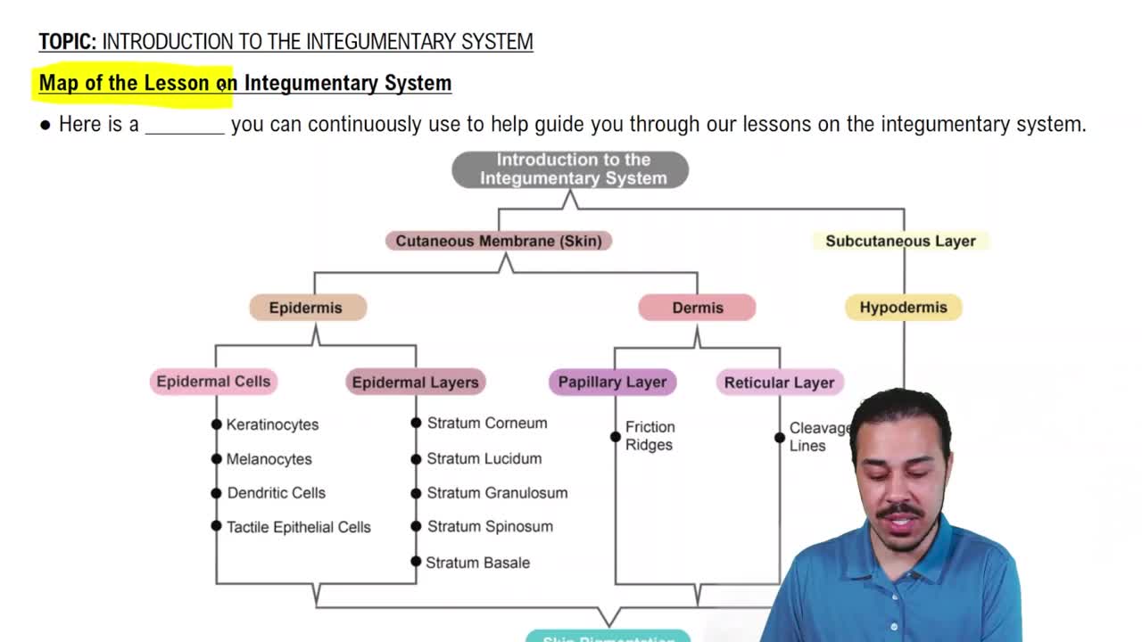
Stratum basale, spinosum, granulosum, lucidum, corneum.
Hemidesmosomes attach basal epidermal cells to the basement membrane; epidermal ridges interlock with dermal papillae to anchor epidermis to dermis.
Basal (stem) cells: produce new keratinocytes.
Melanocytes: synthesize melanin for UV protection.
Tactile (Merkel) cells: sensory for touch.
Due to desmosomes connecting cells with tree-like branching, providing strength and resistance to stress.
Dendritic (Langerhans) cells in the stratum spinosum defend against microorganisms and skin cancer cells.
Born in basale (cell division), keratinization begins in granulosum, cells lose organelles and die, forming dead corneocytes in corneum that are shed continuously.
Insensible: water diffuses across stratum corneum, not felt.
Sensible: water excreted by sweat glands to cool body.
Stimulates epithelial cell growth and repair, including glands in the duodenum and salivary glands.
Papillary: superficial, loose areolar connective tissue, contains capillaries and Meissner's corpuscles.
Reticular: deeper, dense irregular connective tissue with collagen and elastic fibers, provides strength and elasticity.
Collagen fibers provide tensile strength and water-binding; elastic fibers allow recoil and maintain skin turgor.
Parallel cuts align with collagen fibers, keeping wounds closed and reducing scarring; perpendicular cuts pull wounds open.
Dehydration reduces interstitial fluid causing loss of turgor; aging reduces elastic fibers causing loss of recoil and wrinkles.
Merkel: in stratum basale, senses light touch.
Meissner's: in dermal papillae, senses light touch and low-frequency vibration.
Pacinian: deep dermis/subcutaneous, senses deep pressure and vibration.
It lies below the dermis, separating skin from deep fascia; composed mainly of adipose tissue for energy storage and insulation.
Carotene: yellow/orange pigment.
Melanin: brown/tan pigment from melanocytes.
Hemoglobin: red color from blood oxygenation.
Melanocytes package melanin in vesicles (melanosomes) transferred to keratinocytes; more melanosomes produce darker skin.
Sunlight increases melanin production, which protects skin cells from UV-induced DNA damage.
Increased blood flow causes redness; decreased flow and oxygen cause cyanosis (bluish skin); low melanin causes paleness.
Albinism: genetic lack of melanin.
Vitiligo: autoimmune loss of melanin in patches.
Both increase UV susceptibility.
Jaundice: yellowing from bilirubin buildup.
Addison’s: increased ACTH stimulates melanin, darkening skin.
Basal cell: from stratum basale, common, rarely metastasizes.
Squamous cell: from squamous cells, caused by UV, rarely metastasizes.
Melanoma: from melanocytes, aggressive, often metastasizes.
Asymmetry, irregular borders, mottled color, size >6 mm, and changes in size, shape, or color.
Cholecalciferol; produced by epidermal cells stimulated by UV rays.
Liver and kidneys convert cholecalciferol into active hormone calcitriol.
Calcitriol promotes absorption of calcium and phosphate ions in the small intestine.
Hair protects skin from microorganisms; movement sensed by root hair plexus of sensory nerves.
Contracts to pull hair upright, causing goose bumps.