 Back
Anatomy & Physiology: Joint Structure and Movement
 04:06

Terms in this set (80)
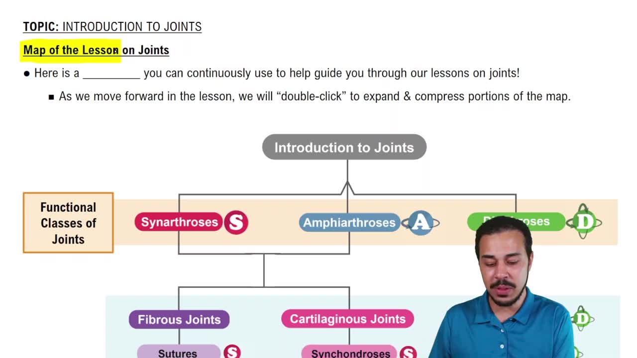
Fibrous, cartilaginous, and synovial.
Dense fibrous connective tissue.
Little to none; many are immovable.
Skull sutures.
Cartilage.
None to slight, depending on the type.
Pubic symphysis and intervertebral discs.
A joint cavity, synovial fluid, and articular cartilage.
Free movement; they are functionally diarthrotic.
Functionally immovable joint.
Functionally slightly movable joint.
Functionally freely movable joint.
Structural classification uses tissues/architecture; functional classification uses amount of movement.
Plane, hinge, pivot, condyloid, saddle, and ball-and-socket.
Mostly flat surfaces; gliding/sliding movement.
Intercarpal joints of the wrist or intertarsal joints of the foot.
Flexion and extension, mainly in one plane.
Elbow (humeroulnar) and interphalangeal joints.
Rotation around one axis.
Atlantoaxial joint (C1-C2) and proximal radioulnar joint.
An oval convex surface fits into an oval depression.
Flexion, extension, abduction, adduction, and circumduction.
Metacarpophalangeal (knuckle) joints and the radiocarpal wrist joint.
Each surface is concave in one direction and convex in the other.
Flexion, extension, abduction, adduction, circumduction, and opposition.
Trapezium and first metacarpal.
A rounded head fits into a cup-like socket.
Flexion, extension, abduction, adduction, rotation, and circumduction.
Shoulder and hip.
Movement around one axis; hinge and pivot joints.
Movement around two axes; condyloid and saddle joints.
Movement around three axes; ball-and-socket joints.
Plane (gliding) joint.
A movement that generally decreases the angle between bones.
A movement that generally increases the angle between bones.
Movement away from the body's midline.
Movement toward the body's midline.
Circular, cone-shaped movement combining flexion, extension, abduction, and adduction.
A bone turns around its longitudinal axis.
Movement of the thumb across the palm toward the fingers.
Humerus, ulna, and radius; the humeroulnar articulation is the main hinge.
The humeral trochlea fits into the ulna's trochlear notch.
The capitulum of the humerus articulates with the head of the radius.
Hinge; allows flexion and extension.
Scapula and humerus.
The head of the humerus fits into the glenoid cavity of the scapula.
Ball-and-socket; multiaxial.
The coxal bone of the pelvis and the femur.
The head of the femur fits into the acetabulum of the pelvis.
Ball-and-socket; multiaxial.
Atlas (C1) and axis (C2).
The dens of C2 rotates within a ring associated with C1 and its transverse ligament.
Pivot; allows rotation (turning the head side to side).
Radius and ulna.
Pivot; permits pronation and supination of the forearm.
Trapezium and first metacarpal.
Saddle; allows thumb opposition.
A metacarpal and the proximal phalanx.
Condyloid; biaxial.
Adjacent phalanges.
Hinge; allows flexion and extension.
Adjacent carpal bones.
Plane; gliding.
An instrument used to measure joint angles and range of motion (ROM).
To assess mobility, record a baseline, and track changes over time.
The joint's axis of rotation.
Along the stationary body segment.
Along the body segment that moves.
Position joint; align fulcrum and arms; move joint; read the angle in degrees.
The amount of movement possible at a joint, usually measured in degrees.
It influences the directions and range of movement possible.
They stabilize joints and limit excessive movement.
They support joints; tightness or muscle tone can limit movement.
A tight capsule can restrict movement; the capsule also stabilizes the joint.
Mobility often decreases with age.
Disuse can cause stiffness and reduced range of motion.
They can improve flexibility and range of motion.
Pain and swelling may reduce movement.
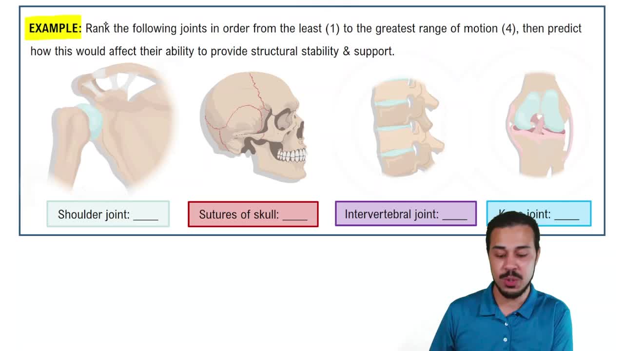
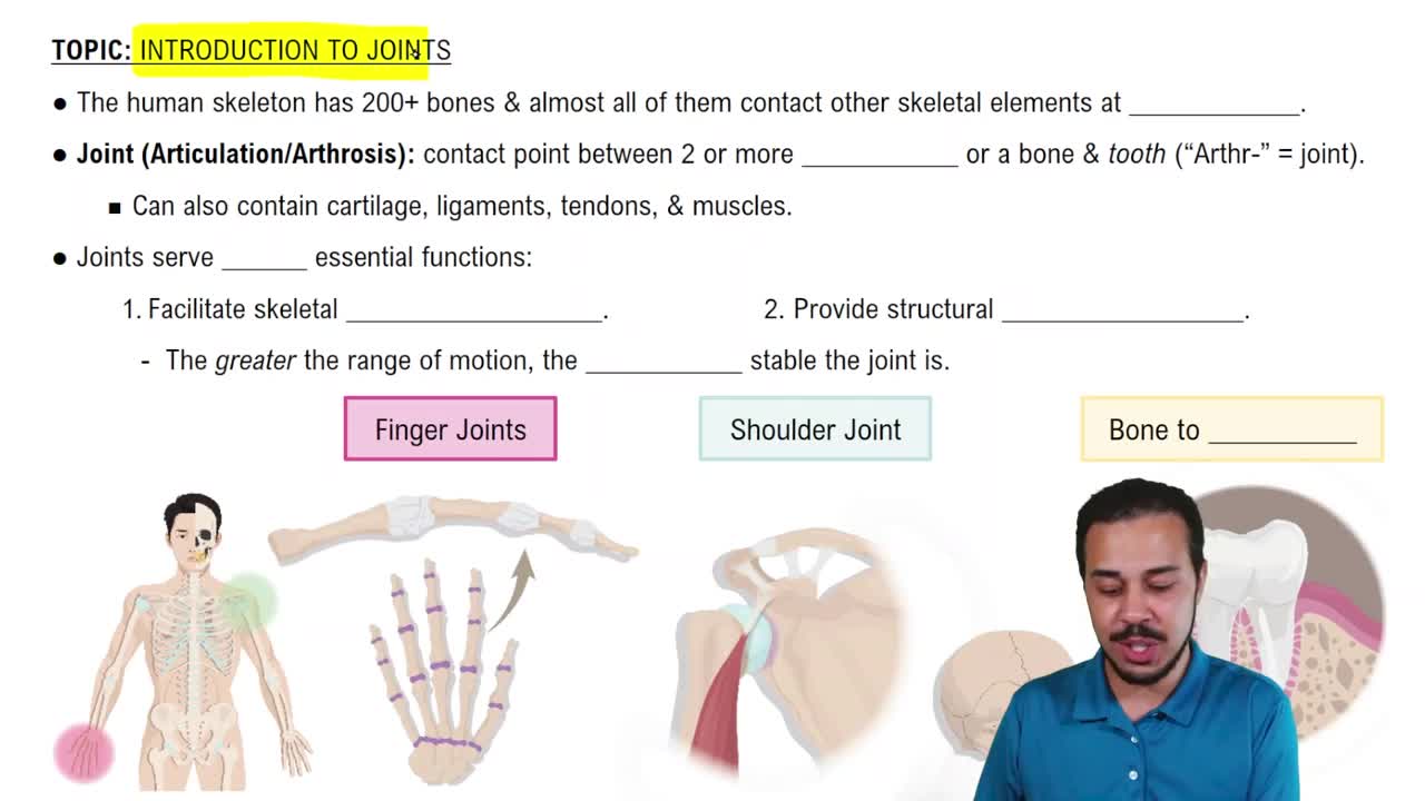
Greater mobility often means less bony stability; stability can limit range of motion.
The shoulder's shallow glenoid allows more movement; the hip's deep acetabulum provides more stability.