 Back
Anatomy & Physiology: Joints & Movement Recall
 04:06

Terms in this set (44)
A synarthrosis is a functionally immovable joint.
Suture (skull), gomphosis (tooth/PDL), synchondrosis (hyaline cartilage bridge), synostosis (bone fusion).
Abduction is movement away from the midline (e.g., fingers: middle finger; toes: 2nd toe).
Adduction is movement toward the midline.
Flexion/extension, abduction/adduction, opposition/reposition, circumduction (combination).
Opposition: thumb touches fingertips; Reposition: thumb returns to anatomical position.
Lateral ankle sprain (ATFL) caused by inversion ± plantarflexion.
Dorsiflexion is toes moving up; main muscle: tibialis anterior.
Plantar flexion is toes moving down; main muscles: gastrocnemius and soleus.
The elbow is a synovial hinge joint (functionally diarthrosis).
An amphiarthrosis is a slightly movable joint.
Triaxial joints move in 3 planes: flexion/extension, abduction/adduction, rotation; example: ball-and-socket joint.
A saddle joint allows biaxial movement; example: thumb 1st carpometacarpal (CMC) joint.
Glide, flexion/extension, abduction/adduction, rotation, circumduction, plus special movements like pronation/supination, dorsiflexion/plantarflexion, inversion/eversion, opposition.
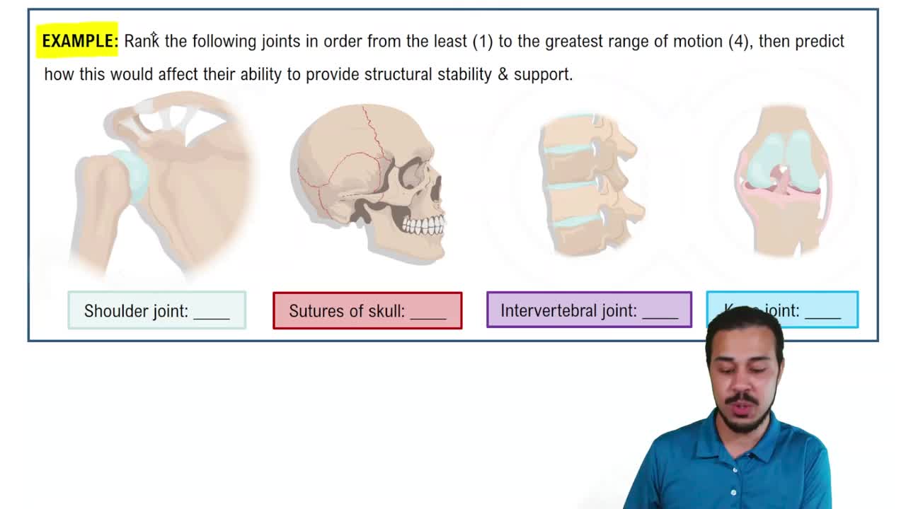
Ball-and-socket joints have the greatest range; the shoulder is the most mobile.
Hyperextension is extending a joint beyond its neutral position; example: neck bending backward past neutral.
Glenohumeral (superior, middle, inferior), coracohumeral, coracoacromial, transverse humeral, acromioclavicular, coracoclavicular (conoid, trapezoid).
The radiocarpal joint is a condyloid/ellipsoid synovial joint, allowing biaxial movement.
Diarthrosis is a freely movable joint, most are synovial joints.
Tear in the annulus fibrosus allows the nucleus pulposus to protrude.
Deep acetabulum, labrum, strong capsule and ligaments, and surrounding muscles.
Iliofemoral, pubofemoral, ischiofemoral, ligamentum teres (± transverse acetabular ligament).
Shock absorption, load distribution, and improving congruency and stability.
The patellar ligament supports the anterior knee.
The oblique popliteal ligament (± arcuate ligament) reinforces the posterior knee.
The medial collateral ligament (MCL/TCL) resists valgus forces.
ACL prevents anterior tibial displacement; PCL prevents posterior tibial displacement.
The ankle is a synovial hinge joint.
The rotator cuff stabilizes the humeral head in the glenoid and aids shoulder motion; muscles: SITS - supraspinatus, infraspinatus, teres minor, subscapularis.
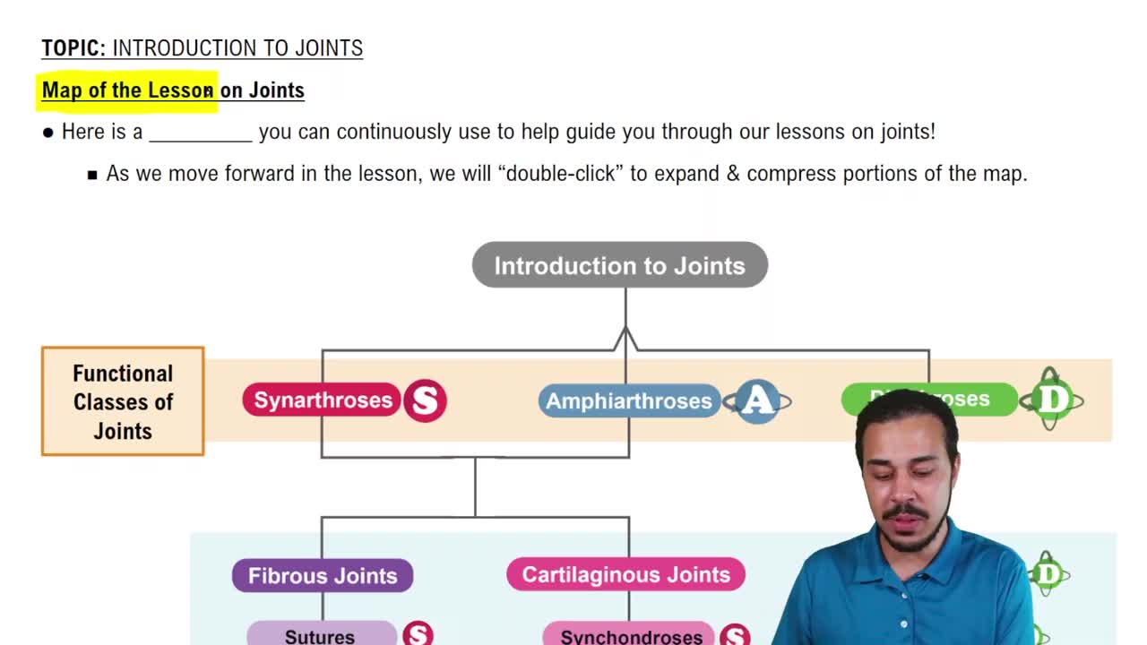
Fibrous, cartilaginous, synovial (classified by binding material and presence of cavity).
Synarthrosis (immovable), amphiarthrosis (slightly movable), diarthrosis (freely movable).
Hyaline cartilage on bone ends; reduces friction, absorbs shock; avascular, so heals slowly.
Eversion is turning the sole of the foot outward.
Supination rotates the forearm so the palm faces up/anterior (like holding soup).
Humeroulnar, humeroradial, and proximal radioulnar joints within one capsule.
Ligaments, tendons, bursae, tendon sheaths, menisci/discs, fat pads, and sometimes labrum.
Lubricates joints, nourishes cartilage, removes waste, reduces friction, and absorbs some shock.
A fibrous joint connected by ligaments or interosseous membrane; example: tibia–fibula.
Provide strong stability with slight movement.
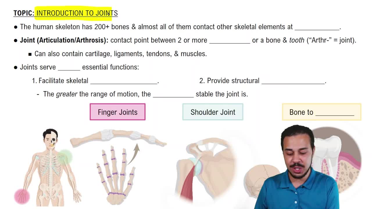
Inverse relationship: more mobility means less stability, and vice versa.
The trochlea is the medial distal humerus that articulates with the ulna.
The femur is the longest and heaviest bone.
The clavicle articulates with the manubrium medially and the acromion laterally.
The synapse between a motor neuron and the muscle fiber's motor end plate; neurotransmitter is acetylcholine (ACh).