 Back
Anatomy & Physiology: Joints and Synovial Joint Features
 04:06

Terms in this set (20)
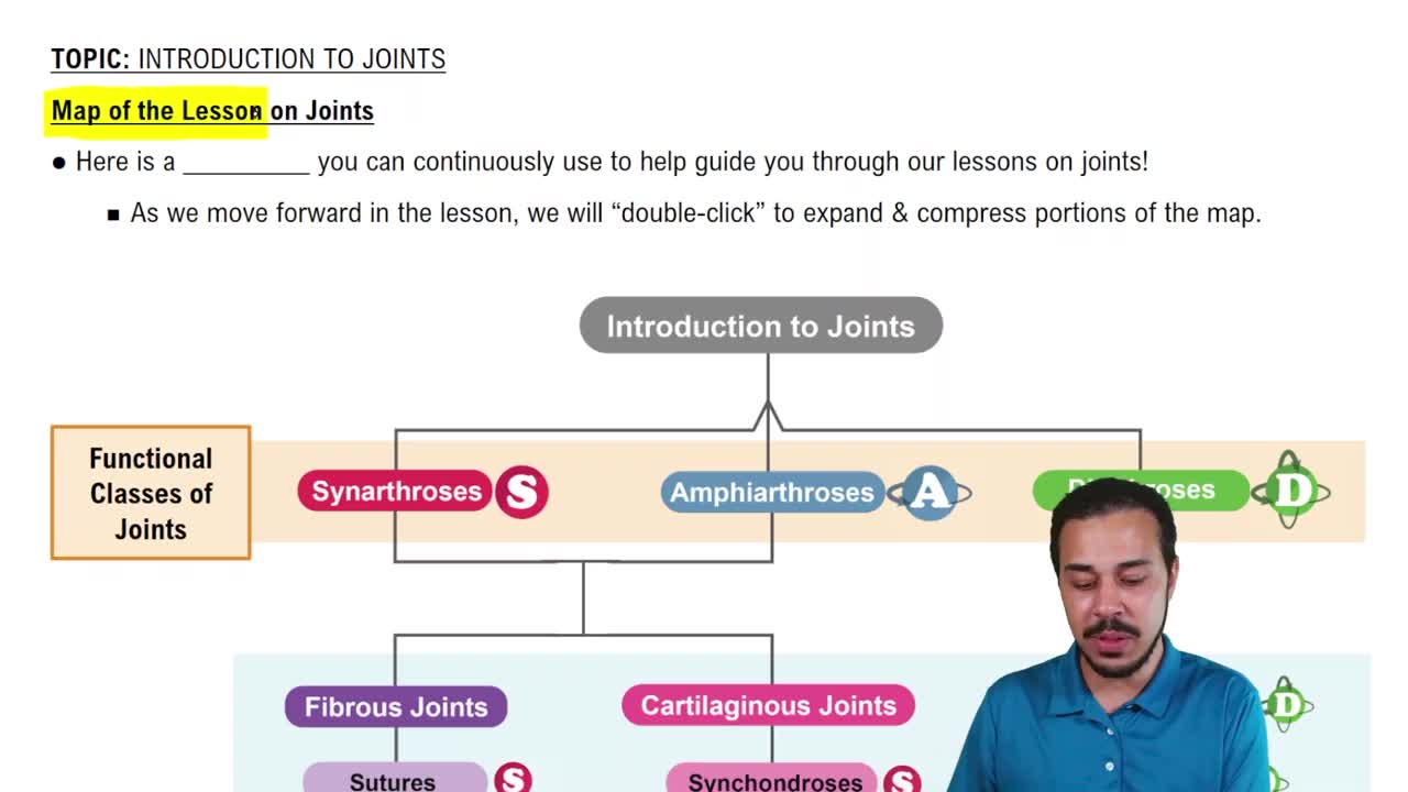
Fibrous, Cartilaginous, and Synovial joints.
Dense fibrous connective tissue joins bones; fibrous joints have no joint cavity.
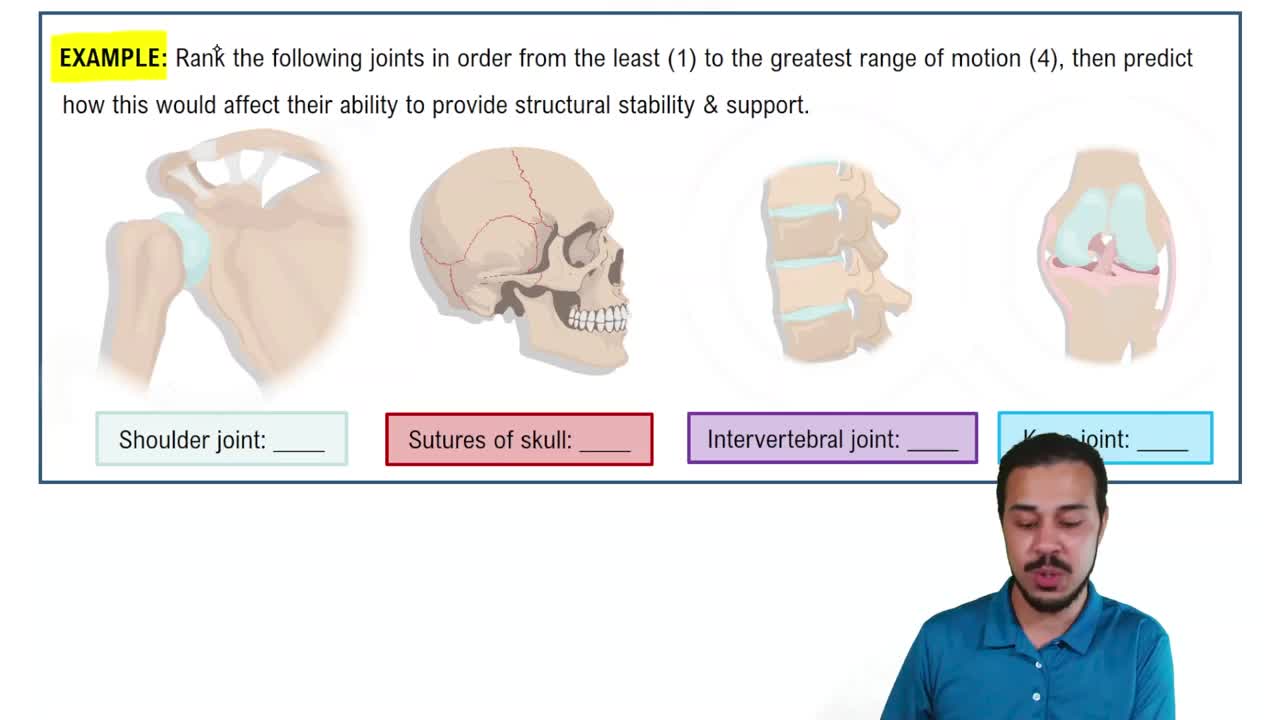
Examples: Sutures. Most fibrous joints are immovable or allow very little movement.
Bones are united by cartilage and have no joint cavity.
Examples: Epiphyseal plates, first rib with sternum, intervertebral joints, pubic symphysis. They are not highly movable.
Synovial joints are freely movable and include almost all limb joints.
1. Articular cartilage
2. Joint (synovial) cavity
3. Articular capsule
4. Synovial fluid
5. Reinforcing ligaments
6. Nerves and blood vessels
Hyaline cartilage covering bone ends that reduces friction and absorbs shock.
A viscous, slippery filtrate of plasma and hyaluronic acid that lubricates and nourishes joint structures.
Bursae are fluid-filled sacs that reduce friction; tendon sheaths are elongated bursae wrapping tendons to reduce friction.
1. Shape of articular surfaces
2. Ligament number and location
3. Muscle tone (most important)
One flat bone surface slides or glides over another similar surface.
Movements that increase or decrease the angle between bones: flexion, extension, hyperextension, abduction, adduction, and circumduction.
Turning a bone around its longitudinal axis, including medial and lateral rotation.
Supination: rotation of radius and ulna to face palm up.
Pronation: rotation to face palm down.
Opposition: thumb movement to touch fingers.
Plane, Hinge, Pivot, Condylar (ellipsoid), Saddle, and Ball-and-socket joints.
A ball-and-socket joint; most freely moving but less stable.
Because the shoulder sacrifices stability for mobility; weakest anteriorly and inferiorly, allowing the humerus to dislocate forward and downward.
Cartilage tears, sprains (ligament stretch/tear), and dislocations (bones forced out of alignment).
Muscle tone keeps tendons taut across joints, reinforcing stability especially in shoulder, knee, and foot arches.