 Back
BackAnatomy & Physiology: Reproductive System
 2:08
2:08
Terms in this set (21)
Spermatogenesis is the process of sperm cell development in the seminiferous tubules, involving mitosis of spermatogonia, meiosis to form spermatids, and spermiogenesis to produce mature sperm.
1. Mitosis of spermatogonia
2. Meiosis producing spermatids
3. Spermiogenesis transforming spermatids into sperm
Sustentocytes support developing sperm cells, form the blood-testis barrier, and regulate the environment for spermatogenesis by controlling substances and protecting haploid cells from immune attack.
By the hypothalamic-pituitary-gonadal (HPG) axis: GnRH from hypothalamus stimulates FSH and LH release from pituitary; LH stimulates testosterone secretion; FSH stimulates sustentocytes to release androgen-binding protein; testosterone and inhibin provide negative feedback.
ABP binds testosterone in the seminiferous tubules, maintaining high local testosterone levels to stimulate spermatogenesis.
High before birth, low during childhood, rises at puberty to adult levels, then remains fairly stable until late in life.
Head: contains nucleus and acrosome for egg penetration.
Midpiece: contains mitochondria for ATP production.
Tail: flagellum for locomotion.
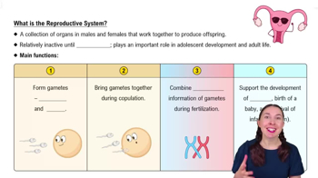
Oogenesis is the production of female gametes (ova), beginning in the fetus with oogonia dividing to form primary oocytes that arrest in prophase I until puberty.
Oogenesis produces one functional ovum plus polar bodies, begins in fetal life and ends at menopause, and has a higher error rate; spermatogenesis produces four sperm continuously from puberty onward.
Ovarian follicles are saclike structures in the ovary cortex containing an immature oocyte surrounded by support cells, maturing through stages from primordial to vesicular follicles.
Several vesicular follicles grow under FSH stimulation; one becomes dominant, and its primary oocyte completes meiosis I to form a secondary oocyte and first polar body.
The mature follicle ruptures, releasing the secondary oocyte with its corona radiata into the peritoneal cavity.
After ovulation, the ruptured follicle forms the corpus luteum, which secretes progesterone and estrogen; if no pregnancy occurs, it degenerates into the corpus albicans.
The uterus receives, retains, and nourishes the fertilized egg; it has three layers: perimetrium, myometrium (muscle), and endometrium (mucosal lining).
Functional layer (stratum functionalis) changes cyclically and is shed during menstruation; basal layer (stratum basalis) is permanent and regenerates the functional layer.
The vagina is a distensible tube with three layers: adventitia, muscularis, and mucosa; it serves as the birth canal, passage for menstrual flow, and copulatory organ.
By interactions of GnRH, FSH, LH, estrogens, and progesterone, which regulate follicle development, ovulation, and uterine changes.
Estrogens promote oogenesis, follicle growth, reproductive tract development, secondary sex characteristics, and a growth spurt.
Progesterone prepares and maintains the uterus for pregnancy and regulates the menstrual cycle during the luteal phase.
Atresia is the programmed cell death of ovarian follicles, the fate of most follicles that do not reach ovulation.
It protects developing sperm cells from immune system attack by isolating haploid cells in the adluminal compartment of seminiferous tubules.