Back

Anatomy & Physiology: Skeletal System and Articulations LAB #5

Control buttons has been changed to "navigation" mode.
1/28
  • Compact bone structure

    Compact bone is arranged in Haversian systems or osteons, with concentric lamellae surrounding a central canal containing blood vessels, nerves, and lymphatics.
  • Osteocytes location

    Osteocytes reside in lacunae at the borders of concentric lamellae and extend cytoplasmic processes through canaliculi.
  • Spongy bone composition

    Spongy bone consists of trabeculae forming a framework with spaces containing red marrow for blood cell production.
  • Periosteum and endosteum

    The periosteum covers the outer bone surface and is anchored by perforating fibers; the endosteum lines the marrow cavity.
  • Axial vs appendicular skeleton

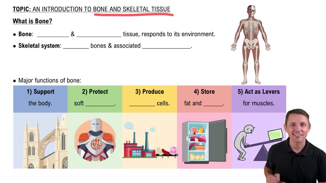
    Axial skeleton includes skull, vertebrae, ribs, and sternum; appendicular skeleton includes girdles and limbs.
  • Cervical, thoracic, lumbar vertebrae count

    Cervical: 7, Thoracic: 12, Lumbar: 5 vertebrae.
  • Sacrum and coccyx

    Sacrum has 5 fused vertebrae articulating with coxal bones; coccyx consists of 2-4 fused vertebrae.
  • Sternum parts

    The sternum consists of the manubrium, body, and xiphoid process.
  • Rib classification

    Ribs 1-7 are true ribs; 8-12 are false ribs; ribs 11 and 12 are floating ribs.
  • Major scapula features

    Acromion, spine, glenoid cavity, and coracoid process are key parts of the scapula.
  • Hand bone groups

    Carpals (8), metacarpals (I-V starting at pollex), and phalanges (proximal, middle, distal, numbered 1-5).
  • Coxal bone composition

    The coxal bone is formed by fusion of ilium, ischium, and pubis.
  • Femur key features

    Greater and lesser trochanters, medial and lateral condyles are important femur landmarks.
  • Types of joints by structure

    Fibrous, cartilaginous, and synovial joints.
  • Functional joint classifications

    Synarthroses (immovable), amphiarthroses (slightly movable), diarthroses (freely movable).
  • Examples of fibrous joints

    Sutures (cranial bones) and gomphoses (tooth sockets).
  • Examples of cartilaginous joints

    Synchondroses (epiphyseal plates) and symphyses (pubic symphysis).
  • Synovial joint subclasses

    Ball and socket, hinge, saddle, ellipsoid, pivot, and gliding joints.
  • Common synovial joint movements

    Abduction/adduction, flexion/extension, rotation, circumduction, supination/pronation, dorsiflexion/plantar flexion.
  • Accessory structures of joints

    Bursae, tendon sheaths, ligaments, tendons, and menisci.
  • Sella turcica location

    A depression in the sphenoid bone housing the pituitary gland.
  • Foramen magnum location

    Large opening in the occipital bone allowing passage of the spinal cord.
  • Lateral malleolus bone

    The lateral malleolus is a feature of the fibula.
  • Trochlea bone

    A pulley-shaped structure on the distal humerus.
  • Cribriform plate bone

    Part of the ethmoid bone, containing foramina for olfactory nerves.
  • Acromion bone

    A bony projection on the scapula forming the highest point of the shoulder.
  • Articular cartilage type

    Hyaline cartilage covers articulating bone surfaces.
  • Function of bursae

    Fluid-filled sacs that reduce friction between tendons and bones or skin.