 Back
Anatomy & Physiology: Skeletal System and Axial Skeleton
 3:56

Terms in this set (24)
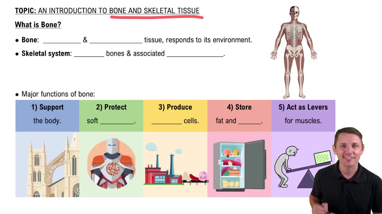
Support, protection, movement, mineral storage, blood cell production, and energy storage.
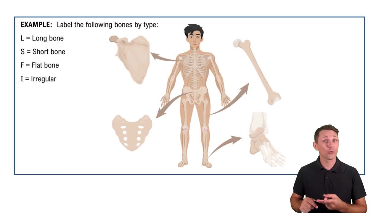
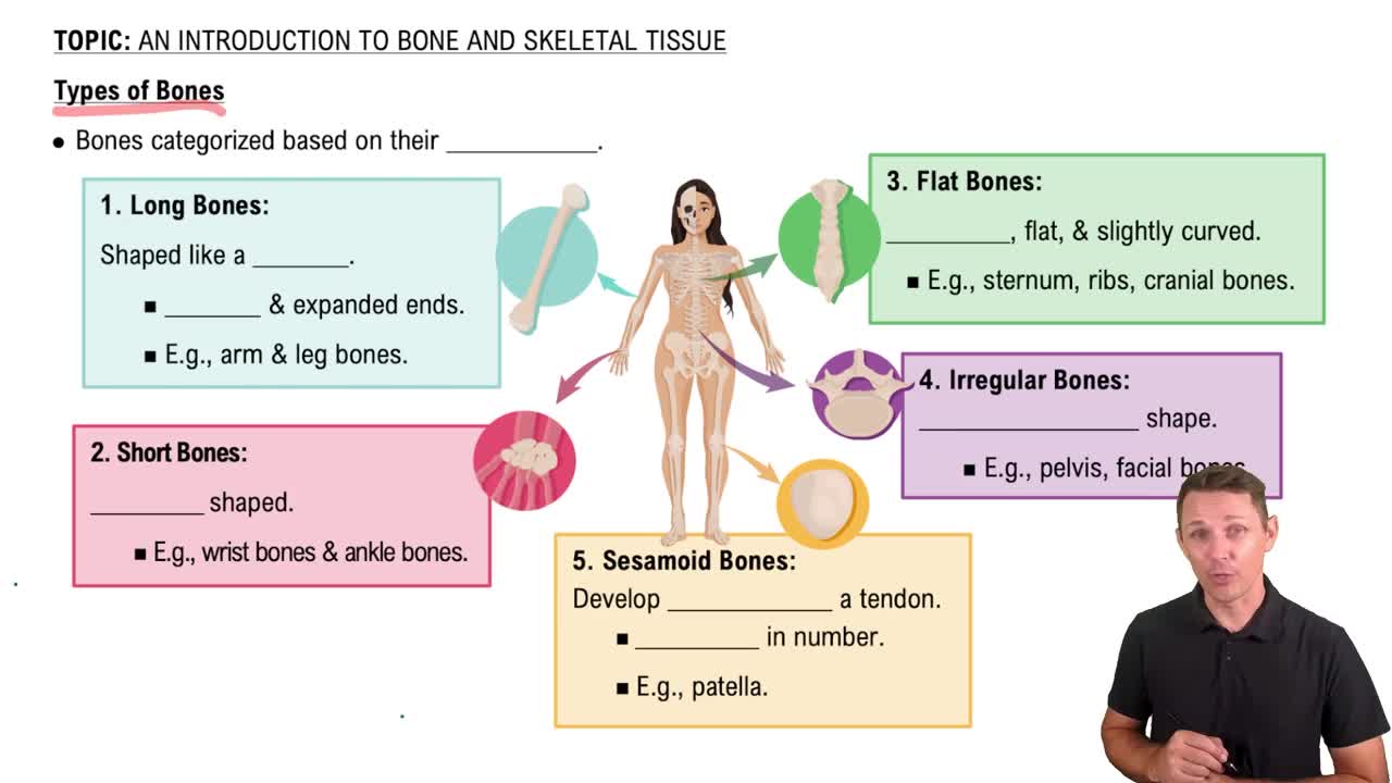
Long bones (e.g., femur), short bones (e.g., carpals), flat bones (e.g., sternum), irregular bones (e.g., vertebrae), and sesamoid bones (e.g., patella).
Bone markings serve as attachment sites for muscles, passageways for nerves and blood vessels, and joint surfaces for articulation.
Osteoblasts build bone, osteocytes maintain bone, osteoclasts resorb bone, and osteoprogenitor cells are stem cells that differentiate into osteoblasts.
Compact bone is dense and provides strength; spongy bone is porous, reduces weight, and contains red marrow for blood cell production.
Bone forms by replacing hyaline cartilage, mainly in long bones during fetal development and growth.
Bone develops directly from mesenchymal tissue, primarily forming flat bones like the skull.
Continuous process of bone resorption by osteoclasts and bone formation by osteoblasts to maintain strength and mineral balance.
Weight-bearing exercise stimulates bone growth and increases bone density.
Calcium is essential for bone strength and mineral storage, and it regulates muscle contraction and nerve function.
Simple, compound, greenstick, comminuted, and spiral fractures, classified by severity and pattern.
Hematoma formation, fibrocartilaginous callus formation, bony callus formation, and bone remodeling.
Decreased bone density, increased fracture risk, reduced calcium absorption, and slower healing.
Includes the skull, vertebral column, and thoracic cage.
Supports and protects the brain, spinal cord, and thoracic organs; provides attachment sites for muscles.
Cranium: frontal, parietal, temporal, occipital. Face: maxilla, mandible, zygomatic.
Allow passage of nerves and blood vessels to and from the brain and face.
Protects the eyes and provides attachment for muscles controlling eye movement.
Lighten the skull, produce mucus, and enhance voice resonance.
Infants have fontanelles and more flexible sutures; adults have fused sutures and fully ossified bones.
Cervical, thoracic, lumbar, and pelvic curves provide balance and absorb shock.
Cervical, thoracic, lumbar, sacral, and coccygeal regions with distinct vertebrae structures.
Allow rib movement for breathing and provide stability to the thoracic cage.
Costosternal joints connect ribs to the sternum, enabling chest expansion during respiration.