 Back
Anatomy & Physiology: Skeletal System and Bone Biology
 3:56

Terms in this set (86)
Axial skeleton and appendicular skeleton.
Approximately 206 bones.
80 bones including the skull, thorax, and vertebral column.
126 bones including limbs and girdles attaching them to the axial skeleton.
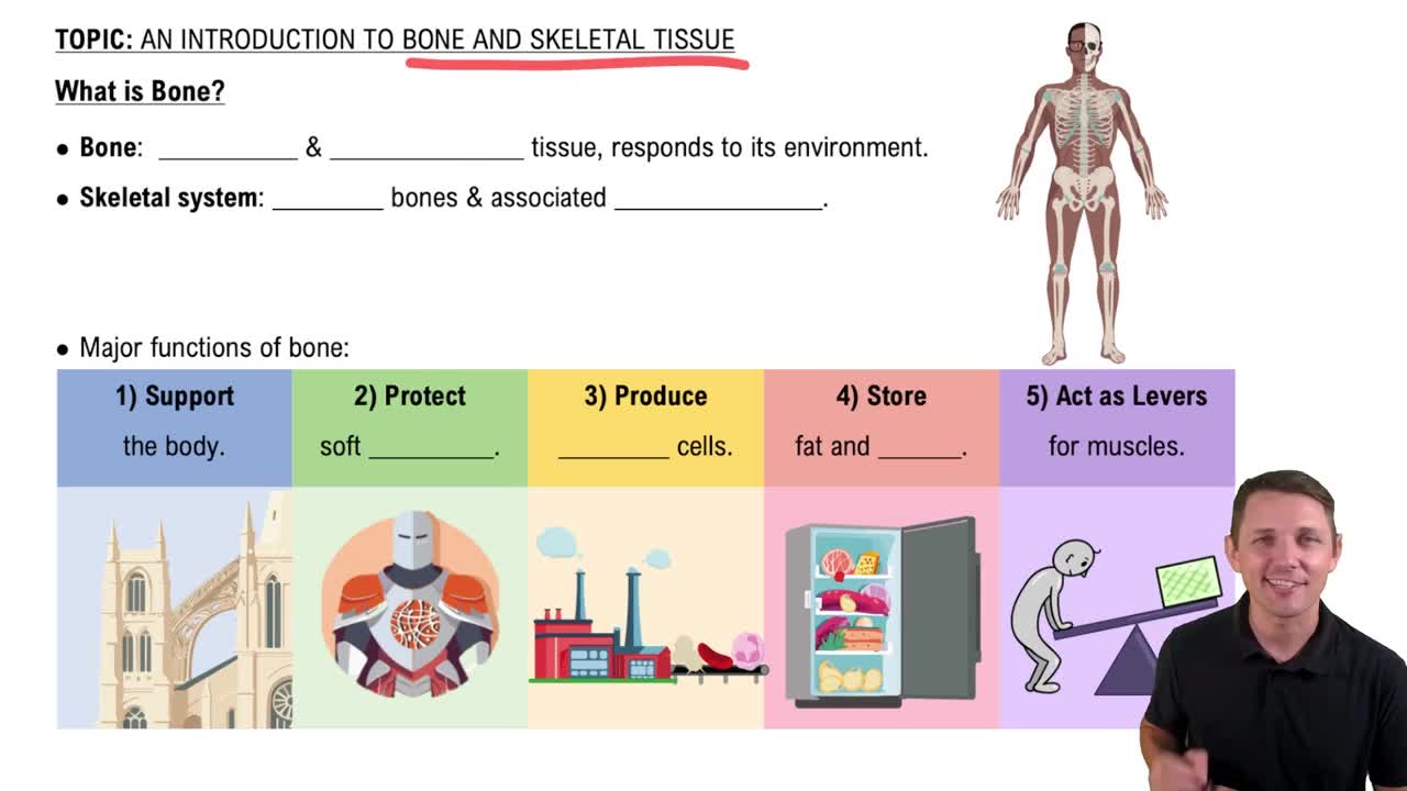
Support the body, anchor muscles, store minerals and fats, produce blood cells in red marrow, protect organs, and act as levers for movement.
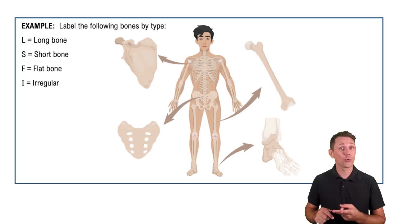
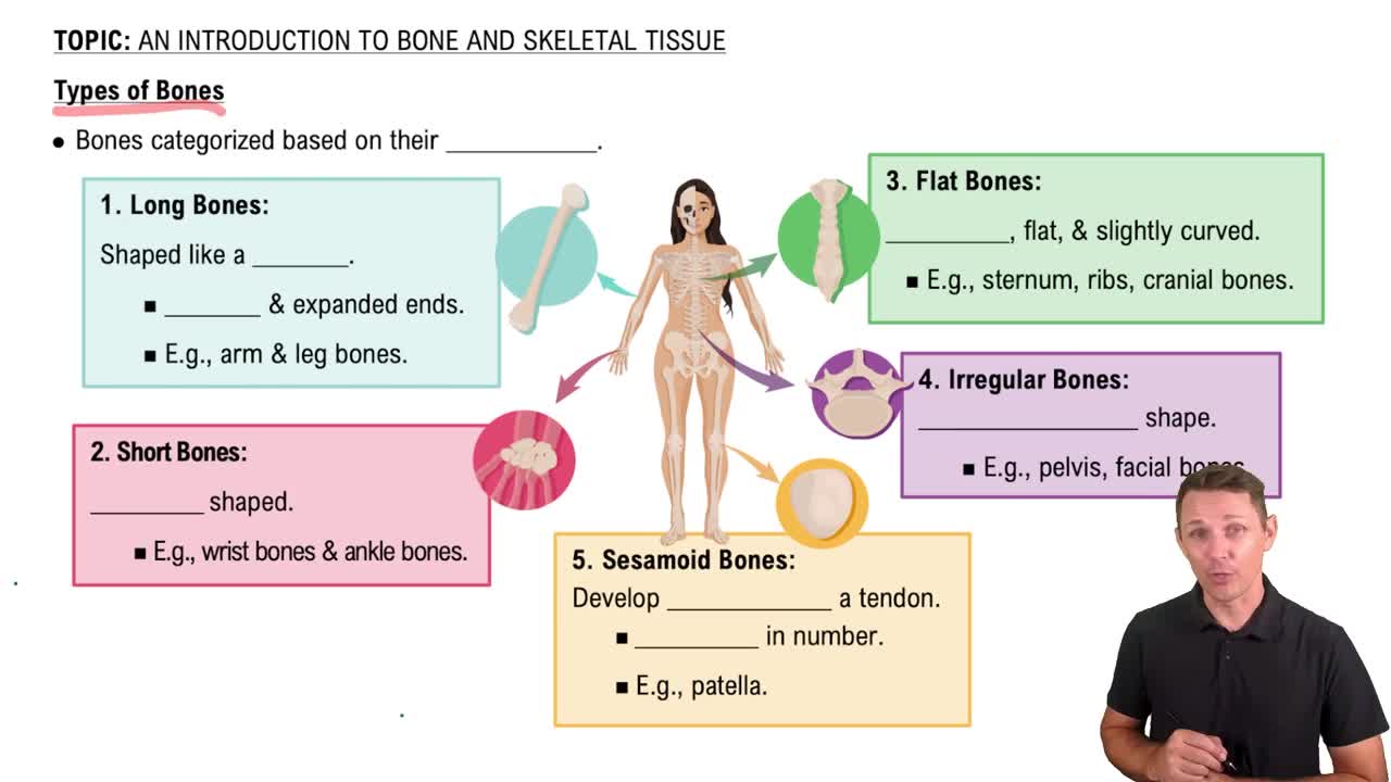
Flat, sutural, long, irregular, sesamoid, and short bones.
Thin bones with parallel surfaces that protect tissues and provide muscle attachment areas.
Cranial bones, sternum, ribs, and scapulae.
Irregular bones forming between cranial bones; their number, size, and shape vary.
Relatively long and slender bones, such as many limb bones.
Bones with complex shapes, e.g., vertebrae, pelvic bones, and facial bones.
Small, flat bones developing within tendons, such as the patella.
Small, box-shaped bones like carpals and tarsals.
The expanded area at each end of a long bone, mostly spongy bone covered by compact bone.
Cartilage covering parts of the epiphysis that form joints.
Region connecting epiphysis to diaphysis; contains the growth plate.
The shaft of a long bone.
The marrow cavity inside the diaphysis of a long bone.
Produces red blood cells, white blood cells, and platelets.
Marrow containing adipose tissue serving as an energy reserve.
A covering around the outer surface of compact bone.
Fibrous outer layer and cellular inner layer.
Separates bone from tissues, provides routes for blood vessels and nerves, and aids bone growth and repair.
Cellular layer lining the medullary cavity, covering spongy bone and central canals.
During bone growth, repair, and remodeling.
An opening allowing blood vessels access to the marrow cavity.
Stem cells producing cells that differentiate into osteoblasts; important in fracture repair.
In the inner periosteum and endosteum lining the medullary cavity.
Cells that produce new bone matrix and help deposit calcium salts.
The unmineralized bone matrix produced by osteoblasts.
The process of forming bone.
Osteocytes.
Mature bone cells maintaining the matrix; they cannot divide.
Small pockets in bone matrix containing osteocytes.
Layers of bone matrix.
Small channels connecting lacunae and the central canal.
Cells that remove and remodel bone matrix by releasing acids and enzymes.
The breakdown of bone matrix releasing stored minerals.
Collagen fibers, about one-third of bone weight.
Calcium phosphate, about two-thirds of bone weight.
Periosteum, compact bone, and spongy bone.
The osteon.
Structural unit of compact bone with concentric lamellae around a central canal.
Channels in osteons containing blood vessels and nerves.
Bone matrix layers at outer and inner surfaces of compact bone.
Bone matrix layers filling spaces between osteons.
Canals connecting blood vessels within compact bone.
Bone with lamellae forming trabeculae, creating an open network.
Between the trabeculae.
Increase in diameter of an existing bone.
Periosteum and endosteum.
Osteoblasts add matrix beneath periosteum; osteoclasts remove matrix from inner surface enlarging medullary cavity.
They mature into osteocytes.
Replacing a cartilage model with bone.
Hyaline cartilage.
Chondrocytes produce cartilage on epiphyseal side; osteoblasts replace cartilage with bone on diaphyseal side.
Central region of cartilage model where bone formation begins in the shaft.
Regions in epiphyses where bone formation begins as capillaries and osteoblasts migrate in.
Cartilage plate separating epiphysis from diaphysis during bone length growth.
Cartilage is replaced by bone, leaving an epiphyseal line.
Bone formation without a prior cartilage model.
Skull roofing bones, lower jaw, collarbone, and sesamoid bones like the patella.
Location where bone formation begins in tissue.
Stem cells cluster, differentiate into osteoblasts, and secrete osteoid that mineralizes.
Osteoblasts become trapped and mature into osteocytes; stem cells produce more osteoblasts.
Remodeling around blood vessels produces osteons of compact bone.
Inorganic ions vital for osmotic balance and physiological processes.
Calcium.
About 99%.
Muscle contraction, blood clotting, and nerve impulse generation.
Absorb calcium and phosphate under hormonal control.
Osteoclasts release calcium by eroding matrix; osteoblasts deposit calcium in new matrix.
Regulate calcium and phosphate loss in urine under hormonal control.
Parathyroid hormone (PTH) and calcitriol.
Promotes bone matrix breakdown by osteoclasts and increases calcium absorption and reabsorption.
Calcitonin.
Inhibits osteoclast activity and promotes calcium deposition in bone matrix.
A crack or break caused by extreme mechanical load.
A fracture that remains internal with no break in the skin.
A fracture projecting through the skin, increasing infection and bleeding risks.
Fracture hematoma formation, callus formation, spongy bone formation, and compact bone formation.
A large blood clot closing injured vessels, developing within hours after fracture.
Network of spongy bone uniting inner fracture edges.
Cartilage and bone structure stabilizing outer fracture edges.
Cartilage of external callus is replaced by spongy bone; dead bone and fragments are removed.
Spongy bone is replaced by compact bone; remodeling reduces fracture evidence.