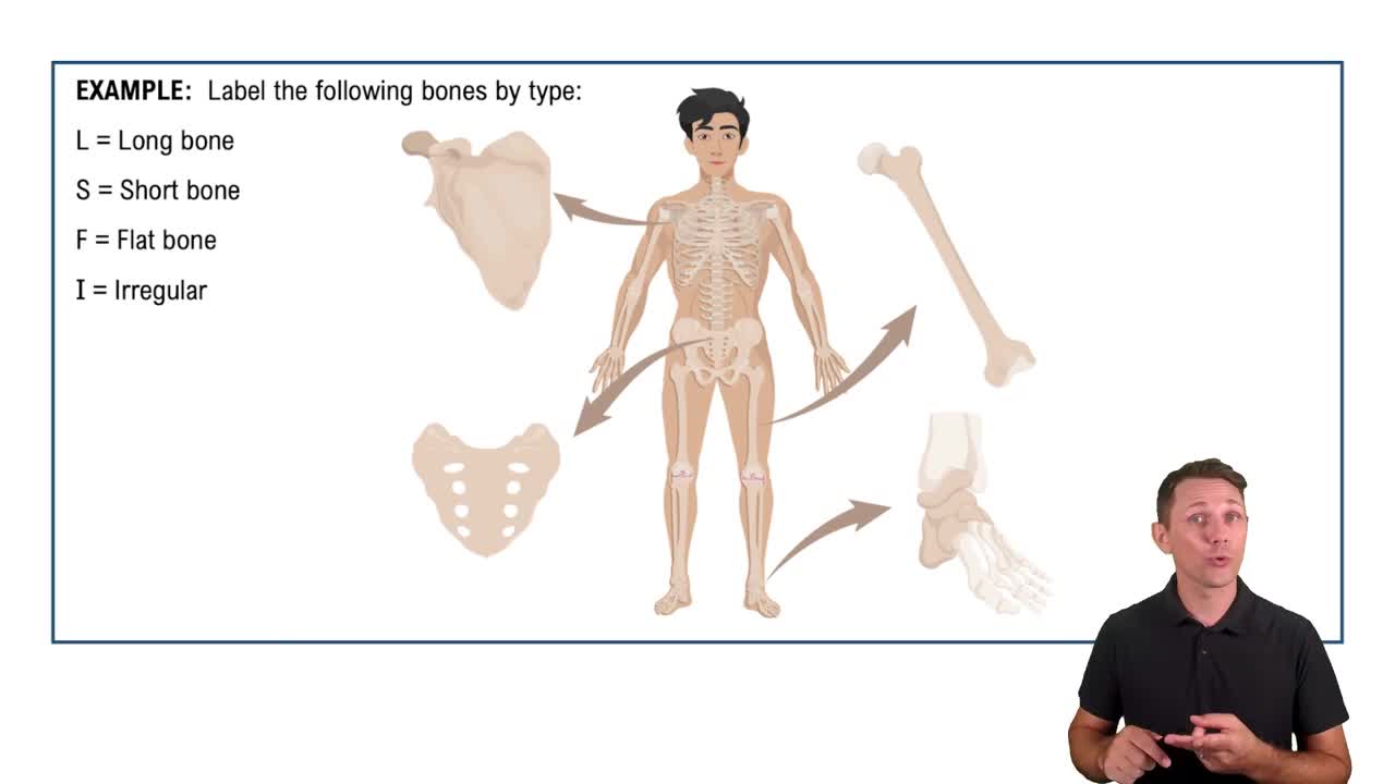
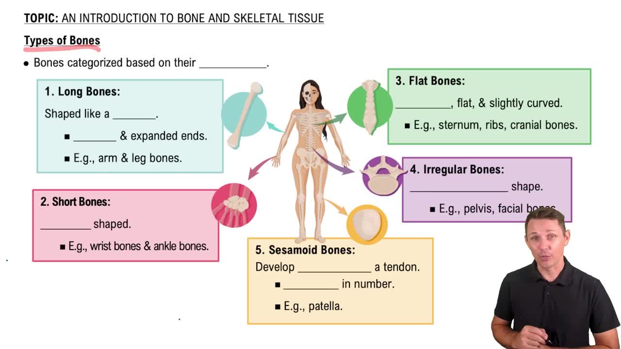
Long bones are longer than wide; short bones are cube-shaped; flat bones are thin and flat; irregular bones have complex shapes; sesamoid bones develop within tendons.
Intramembranous vs. Endochondral bone
Intramembranous bone forms directly from mesenchyme; endochondral bone forms by replacing hyaline cartilage.
Compact bone vs. Spongy bone
Compact bone is dense and forms the outer layer; spongy bone is porous and found inside bones, containing trabeculae.
Periosteum
A dense connective tissue membrane covering the outer surface of bones, containing blood vessels and nerves.
Endosteum
A thin membrane lining the marrow cavity and inner surfaces of bones.
Haversian system (osteon)
The structural unit of compact bone, consisting of a central canal surrounded by concentric lamellae.
Osteocyte
A mature bone cell located in lacunae that maintains bone tissue.
Osteoblast
A bone-forming cell that secretes the bone matrix.
Osteoclast
A large cell that breaks down bone tissue during remodeling.
Lamellae
Concentric rings of calcified matrix in compact bone.
Canaliculi
Tiny channels connecting lacunae, allowing osteocytes to communicate and exchange nutrients.
Epiphyseal plate
A hyaline cartilage plate in the metaphysis where bone growth occurs in children and adolescents.
Intramembranous ossification
Bone formation directly from mesenchymal tissue, forming flat bones like the skull.
Endochondral ossification
Bone formation by replacing hyaline cartilage, forming most long bones.
Functions of the skeletal system
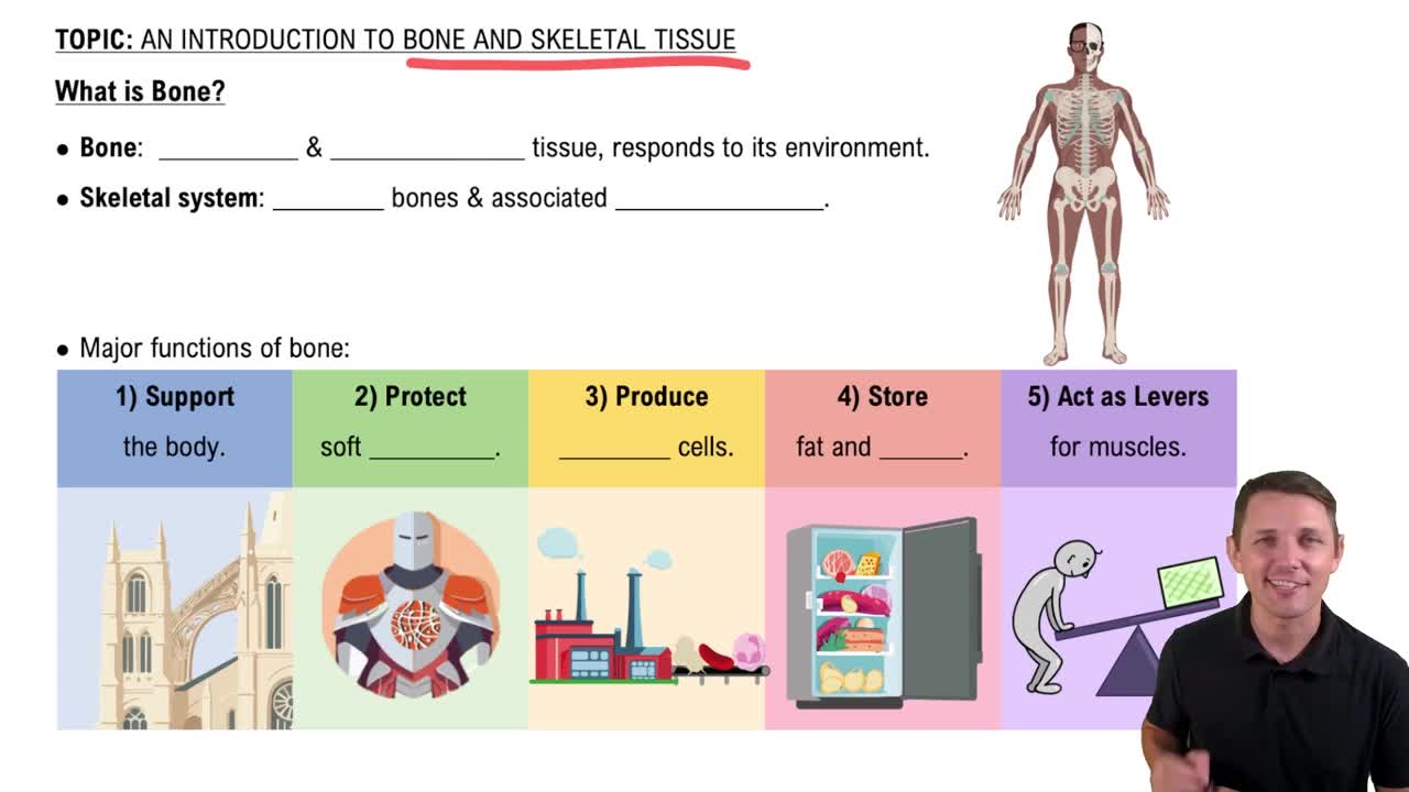
Support, protection, movement, mineral storage, and blood cell production.
Bone remodeling homeostasis
Balance between osteoblast bone formation and osteoclast bone resorption to maintain bone strength and calcium levels.
Parathyroid hormone (PTH) role
Increases blood calcium by stimulating osteoclasts and enhancing calcium absorption in kidneys and intestines.
Calcitonin role
Lowers blood calcium by inhibiting osteoclast activity and promoting calcium deposition in bone.
Bone fracture repair steps
Hematoma formation, fibrocartilaginous callus formation, bony callus formation, and bone remodeling.
Axial vs. Appendicular skeleton
Axial skeleton includes skull, vertebral column, and rib cage; appendicular skeleton includes limbs and girdles.
Cervical, thoracic, lumbar, sacral curves
Cervical and lumbar are concave posteriorly; thoracic and sacral are convex posteriorly, providing balance and flexibility.
Fontanelles in newborn skull
Soft spots: anterior, posterior, sphenoidal, and mastoid fontanelles allow skull growth and passage through birth canal.
Male vs. female pelvis differences
Female pelvis is wider, shallower, and has a larger pelvic inlet for childbirth; male pelvis is narrower and taller.
Bone markings: process, tubercle, condyle
Process is a projection; tubercle is a small rounded projection; condyle is a rounded articular surface.
Marrow cavity
Central cavity of long bones containing bone marrow, which produces blood cells.
Spongy bone histology
Consists of trabeculae with spaces filled by marrow; lacks osteons found in compact bone.
Sutures of the skull
Immovable joints between skull bones: sagittal, coronal, lambdoidal, and squamous sutures.
 Back
 3:56