Skip to main content
Anatomy & Physiology
My Course
Learn
Exam Prep
AI Tutor
Study Guides
Textbook Solutions
Flashcards
Explore
Try the app
My Course
Learn
Exam Prep
AI Tutor
Study Guides
Textbook Solutions
Flashcards
Explore
Try the app
Back
Anatomy & Physiology: Skeletal System and Vertebral Column
You can tap to flip the card.
Bone classification by shape
You can tap to flip the card.
👆
Bone classification by shape
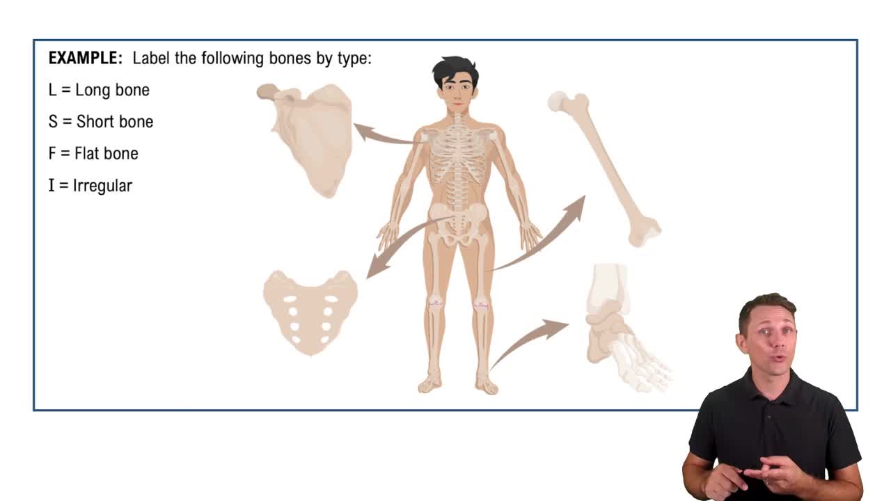
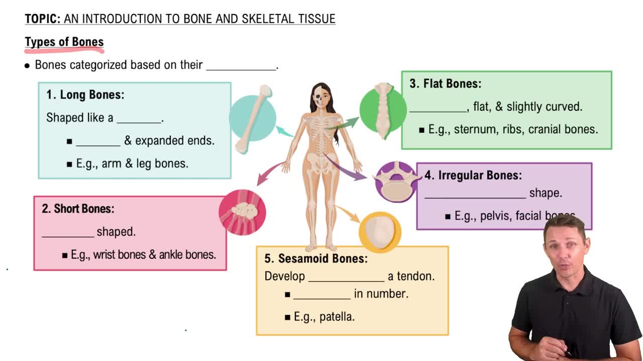
Bones are classified by shape into categories such as long, short, flat, irregular, and sesamoid bones.
Track progress
Control buttons has been changed to "navigation" mode.
1/29
Recommended videos
Guided course
3:56
What is Bone?
29695
views
936
rank
3
comments
Guided course
3:00
An Introduction to Bone and Skeletal Tissue Example 2
16623
views
357
rank
1
comments
Guided course
4:14
Types of Bones
19744
views
503
rank
1
comments
Terms in this set (29)
Hide definitions
Bone classification by shape
Bones are classified by shape into categories such as long, short, flat, irregular, and sesamoid bones.
Sesamoid bones
Small bones resembling sesame seeds, embedded within tendons; the patella is a large sesamoid bone shaped like a shallow disk.
Appendicular skeleton
Includes bones of the upper and lower limbs (appendages).
Axial skeleton
Consists of bones forming the vertical axis: skull, vertebral column, ribs, and sternum.
Compact bone
Dense bone tissue forming the hard exterior covering of all bones.
Spongy (cancellous) bone
Porous bone tissue inside bones, made of trabeculae with spaces filled by red or yellow bone marrow.
Medullary cavity
Hollow space inside the diaphysis of long bones, filled with yellow marrow in adults for fat storage.
Diploë
Spongy bone layer sandwiched between two thin layers of compact bone in flat bones like the parietal bone.
Red bone marrow location in adults
Found in spongy bone of ribs, sternum, cranial bones, vertebrae, clavicles, scapulae, pelvic bones, and proximal epiphyses of long bones.
Yellow marrow function
Stores fat and can convert to red marrow during severe blood loss to increase blood cell production.
Cranial bones
Eight bones forming the walls, roof, and floor of the cranial cavity housing the brain; mostly flat or slightly curved.
Facial bones
Fourteen irregular bones forming the face, orbital and nasal cavities, and bony sockets for teeth.
Bony orbit composition
Formed by seven bones: frontal, ethmoid, lacrimal, sphenoid, maxilla, zygomatic, and palatine.
Fontanelles in fetal skull
Soft spots where cranial bones are connected by fibrous tissue; include anterior, posterior, sphenoidal, and mastoid fontanelles.
Primary curvatures of vertebral column
Thoracic and sacral curvatures, concave anteriorly, present from fetal life.
Secondary curvatures of vertebral column
Cervical and lumbar curvatures, concave posteriorly, develop during infancy and childhood.
Kyphosis
Exaggerated thoracic curvature causing a hunchback appearance.
Lordosis
Exaggerated lumbar curvature resulting in a swayback appearance.
Scoliosis
Abnormal lateral deviation of the vertebral column.
Vertebral body
Anterior bony mass of a vertebra that transmits body weight; increases in size from superior to inferior.
Spinous and transverse processes
Spinous process projects posteriorly; two transverse processes project laterally from each vertebra.
Intervertebral foramina
Openings between adjacent vertebrae allowing spinal nerves to exit the vertebral canal.
Intervertebral discs
Fibrocartilage pads separating vertebral bodies, absent between C1-C2 and fused sacral/coccygeal vertebrae.
Cervical vertebrae features
Smallest vertebrae with transverse foramina for vertebral arteries; C3-C6 have bifid spinous processes; C7 has a prominent non-bifid spinous process.
Atlas (C1)
Ringlike vertebra lacking body and spinous process; articulates with occipital condyles to allow 'yes' head nodding.
Axis (C2)
Has dens projecting superiorly into atlas, forming atlanto-axial joint for 'no' head rotation.
Thoracic vertebrae characteristics
Articulate with ribs via costal facets; bodies are heart-shaped; spinous processes project inferiorly.
Rib articulation with vertebrae
Most ribs articulate with two thoracic vertebrae and the intervertebral disc between them.
Typical rib anatomy
Vertebral end has head, neck, and tubercle; shaft has an angle and costal groove for vessels and nerves.
 BackBack
 3:56