 Back
Anatomy & Physiology: Skeletal System Essentials
 3:56

Terms in this set (20)
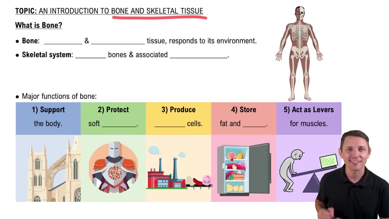
Support, protection, movement, mineral storage, blood cell production, and energy storage.
The outer fibrous layer provides protection and attachment for tendons and ligaments; the inner cellular layer contains osteogenic cells for bone growth and repair.
Cartilage is a flexible connective tissue with no blood vessels, composed of chondrocytes in a matrix of collagen and proteoglycans.
Hyaline, elastic, and fibrocartilage, each differing in fiber composition and location.
Found on articular surfaces of bones, the nose, trachea, and larynx.
Located in the external ear and epiglottis, providing flexibility.
Found in intervertebral discs, pubic symphysis, and menisci of the knee, providing strong support and shock absorption.
Osteoblasts build bone, osteocytes maintain bone, osteoclasts resorb bone, and osteogenic cells are stem cells that differentiate into osteoblasts.
Includes the diaphysis (shaft), epiphyses (ends), medullary cavity, periosteum, and endosteum.
Compact bone is dense and forms the outer layer; spongy bone is porous and found inside bones, especially at the epiphyses.
Forms the outer shell of the diaphysis and epiphyses, providing strength and protection.
Found mainly in the epiphyses and lining the medullary cavity, containing red bone marrow.
Organized into osteons with central canals, concentric lamellae, lacunae with osteocytes, and canaliculi.
Consists of a network of trabeculae with spaces filled by bone marrow.
Responsible for bone formation by secreting osteoid and initiating mineralization.
Maintain bone tissue by regulating mineral content and communicating with other bone cells.
Break down bone matrix during bone resorption to release minerals into the blood.
Stem cells that differentiate into osteoblasts, aiding in bone growth and repair.
Provides a surface for muscle attachment, contains blood vessels and nerves, and supports bone growth and repair.
Houses yellow bone marrow in adults, which stores fat, and in children contains red marrow for blood cell production.