Skip to main content
Anatomy & Physiology
My Course
Learn
Exam Prep
AI Tutor
Study Guides
Textbook Solutions
Flashcards
Explore
Try the app
My Course
Learn
Exam Prep
AI Tutor
Study Guides
Textbook Solutions
Flashcards
Explore
Try the app
Back
Anatomy & Physiology: Skeletal System Joints
You can tap to flip the card.
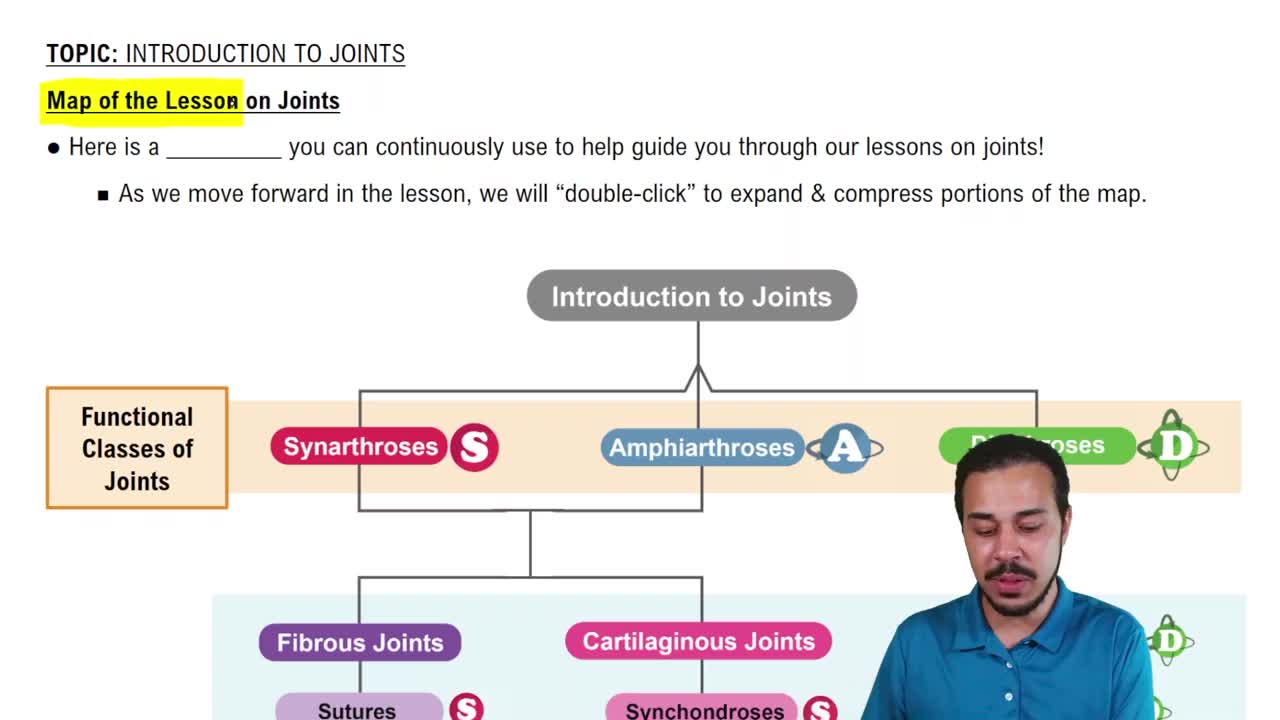
What are the three functional classifications of joints based on range of motion?
You can tap to flip the card.
👆
What are the three functional classifications of joints based on range of motion?
Synarthrosis
(immovable),
Amphiarthrosis
(slightly movable), and
Diarthrosis
(freely movable).
Track progress
Control buttons has been changed to "navigation" mode.
1/26
Recommended videos
Guided course
04:06
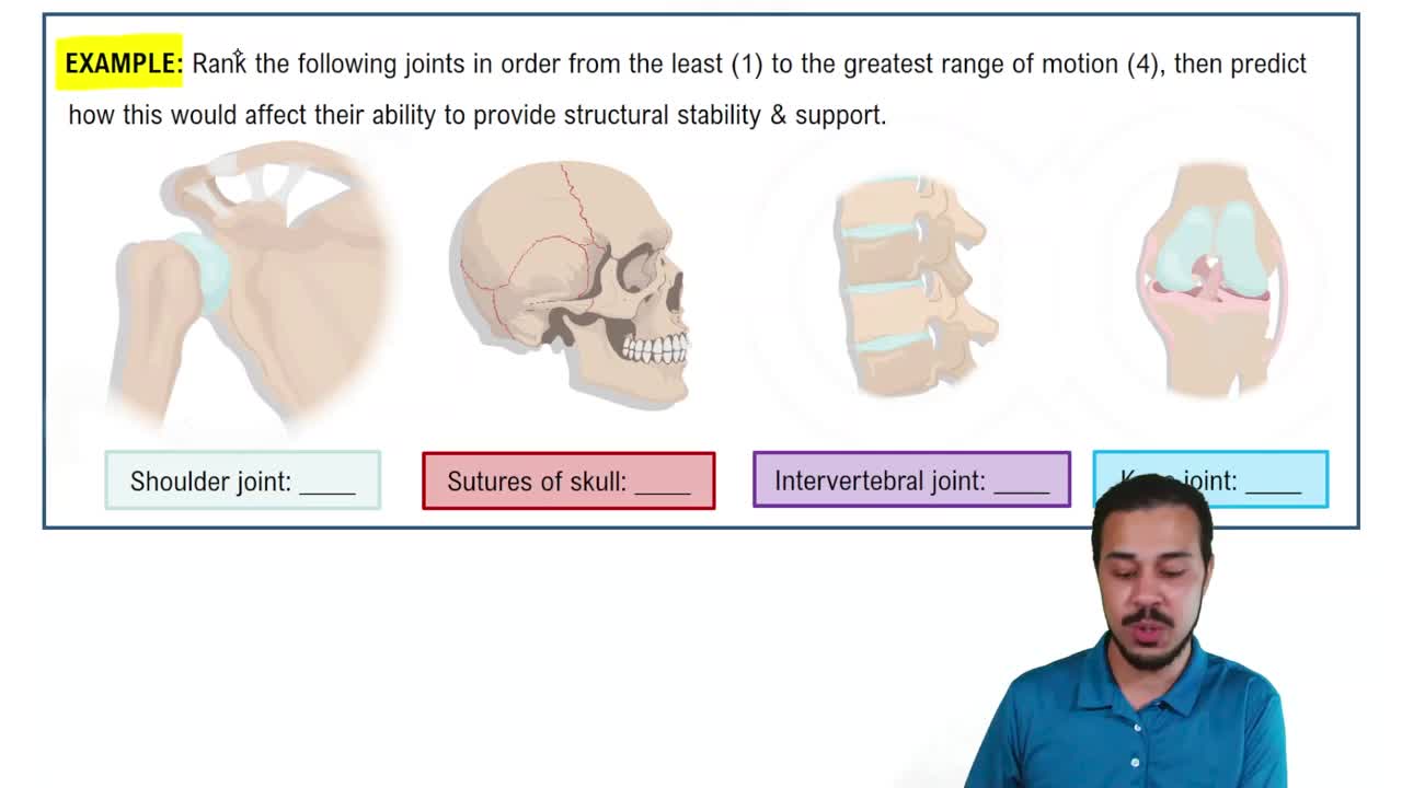
Introduction to Joints Example 1
11986
views
269
rank
Guided course
05:29
Map of the Lesson on Joints
12513
views
264
rank
1
comments
Guided course
05:39
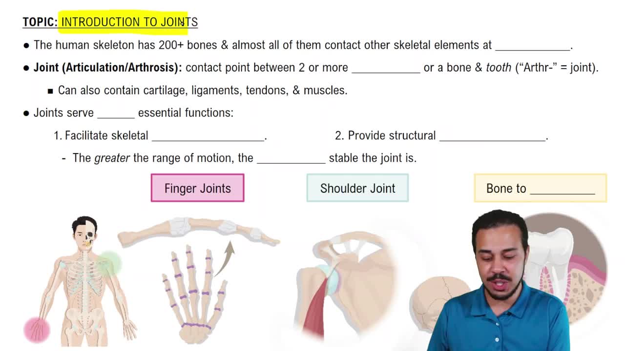
Introduction to Joints
16793
views
431
rank
Terms in this set (26)
Hide definitions
What are the three functional classifications of joints based on range of motion?
Synarthrosis
(immovable),
Amphiarthrosis
(slightly movable), and
Diarthrosis
(freely movable).
What structural types of joints are classified as synarthrosis (immovable)?
Sutures, gomphosis, synchondrosis, and synostosis.
What is a gomphosis joint?
A fibrous joint between teeth and jaw bones held by periodontal ligaments.
What is a synchondrosis joint?
A cartilaginous joint within the epiphysis of a bone that binds the diaphysis to the epiphysis.
What is synostosis?
A bony fusion joint where two bones fuse, such as the fusion of the three coxal bones.
What are amphiarthroses joints and examples?
Slightly movable joints like syndesmosis (ligaments connecting radius and ulna) and symphysis (cartilage pad between pubic bones).
What defines a diarthrosis joint?
A freely movable synovial joint typically found at the ends of long bones, e.g., shoulder, elbow, hip, and knee joints.
Name the six basic characteristics of synovial joints.
Joint capsule, articular cartilages, joint cavity with synovial fluid, synovial membrane, accessory structures (cartilage, ligaments, tendons, bursae), sensory nerves and blood vessels.
What is the function of synovial fluid?
Lubricates articular cartilage surfaces, nourishes chondrocytes, and acts as a shock absorber.
What accessory structures are found in synovial joints?
Cartilages (menisci), fat pads, ligaments (extracapsular and intracapsular), tendons, bursae, and synovial tendon sheaths.
How does joint mobility relate to joint strength?
Highly mobile joints (diarthrosis) are less strong; immovable joints (synarthrosis) are stronger and less prone to injury.
List the types of angular motion at synovial joints.
Abduction, adduction, flexion, extension, hyperextension, and circumduction.
What is rotation in joint movement?
Movement around a longitudinal axis, including left/right rotation, medial/lateral rotation, pronation, and supination.
Name some special movements of synovial joints.
Inversion/eversion, dorsiflexion/plantar flexion, lateral flexion, protraction/retraction, opposition/reposition, elevation/depression.
What bones form the temporomandibular joint (TMJ)?
The condylar process of the mandible and the mandibular fossa of the temporal bone.
Name the ligaments associated with the TMJ.
Lateral ligament, stylomandibular ligament, and sphenomandibular ligament.
What are the components of intervertebral discs?
Anulus fibrosus (outer layer), nucleus pulposus (inner layer), and vertebral endplates.
What ligaments support the vertebral column?
Anterior longitudinal, posterior longitudinal, supraspinous, interspinous, and ligamentum flavum.
What movements occur at the vertebral column?
Flexion, extension, lateral flexion, and rotation.
What bones and joints make up the shoulder complex?
Clavicle, scapula, humerus, sternoclavicular joint, and shoulder (glenohumeral) joint.
What ligaments stabilize the sternoclavicular joint?
Anterior and posterior sternoclavicular ligaments, interclavicular ligament, and costoclavicular ligament.
What type of joint is the shoulder (glenohumeral) joint?
A ball-and-socket joint formed by the head of the humerus and the glenoid cavity.
Name key ligaments of the shoulder joint.
Glenohumeral, coracohumeral, coraco-acromial, acromioclavicular, coracoclavicular, and transverse humeral ligaments.
What is the role of the rotator cuff muscles?
They stabilize the shoulder joint more than the ligaments.
What bursae are associated with the shoulder joint?
Subacromial, subcoracoid, subdeltoid, and subscapular bursae.
How does aging affect joints?
Joints experience wear and tear leading to rheumatism and arthritis, which damage articular cartilage.
 BackBack
 04:06