 Back
Anatomy & Physiology: Skin Structure and Function
 06:54

Terms in this set (30)
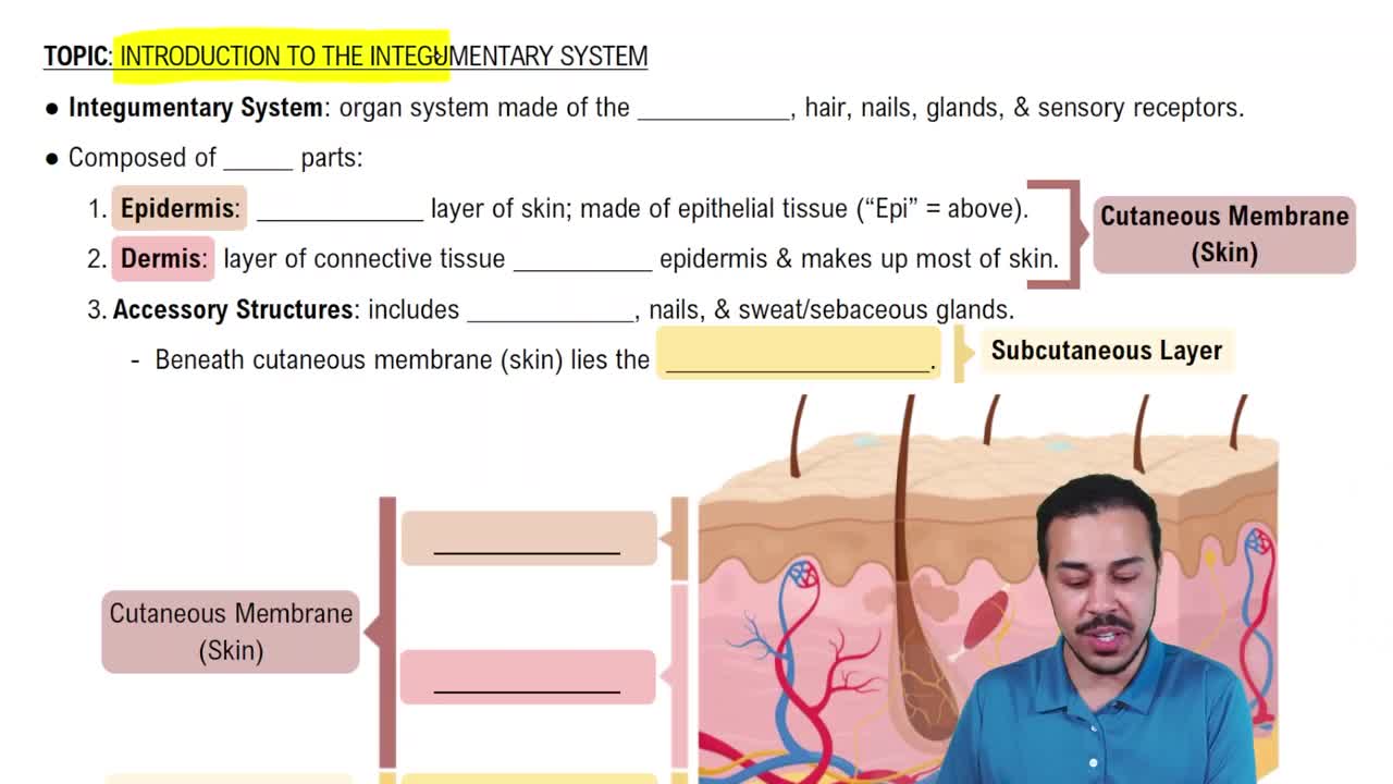
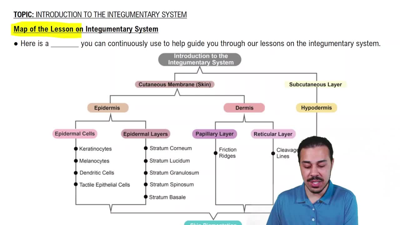
Epidermis, Dermis, and Hypodermis.
Mostly stratified squamous epithelium.
By diffusion from blood vessels in the underlying dermis.
Keratinocytes, Melanocytes, Langerhans cells (immune activators), and Merkel cells (sensory receptors).
They produce keratin, which provides the skin with its protective, waterproof properties.
Melanocytes produce melanin, which protects against UV damage and determines skin color.
They have more active melanocytes producing more melanin, which is distributed differently in keratinocytes.
It is the deepest epidermal layer where cell division (mitosis) produces new keratinocytes.
Keratinization begins in the stratum spinosum.
Desmosomes are cell junctions found in the epidermis that provide mechanical strength by linking keratinocytes.
The stratum lucidum is found only in thick skin like palms and soles.
It consists of dead, keratinized cells that form a protective barrier against environmental damage and water loss.
Loose areolar connective tissue.
Finger-like projections of the dermis that increase surface area and interlock with the epidermis.
They are sensory receptors in the dermal papillae that detect light touch.
It consists of dense irregular connective tissue with collagen and elastin fibers providing strength and elasticity.
They are natural tension lines in the dermis formed by collagen fiber orientation, important for surgical incisions.
Rapid stretching of skin tears the dermis, damaging collagen and elastin fibers.
Mostly adipose tissue that provides insulation and cushioning.
Blood flow in dermal capillaries, especially oxygenated hemoglobin.
Low oxygen levels in blood cause a bluish tint.
Increased melanin production stimulated by excess ACTH.
It causes DNA mutations and can lead to skin cancer.
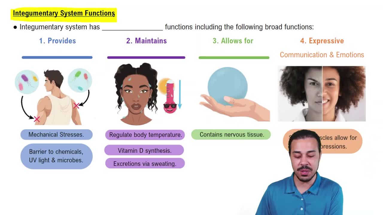
Protection, sensory input, and regulation of body temperature.
It is the outermost layer of the hair shaft, composed of overlapping keratinized cells.
Loss of pigment production by melanocytes in hair follicles.
Eccrine glands (thermoregulation) and apocrine glands (active after puberty, produce odor).
Blocked sebaceous glands and bacterial infection leading to inflammation.
Asymmetry, Border irregularity, Color variation, Diameter over 6 mm.
First degree: epidermis only; Second degree: epidermis and part of dermis; Third degree: full thickness including hypodermis.