 Back
BackAnatomy & Physiology: Skin, Tissues, and Glands
 06:54
06:54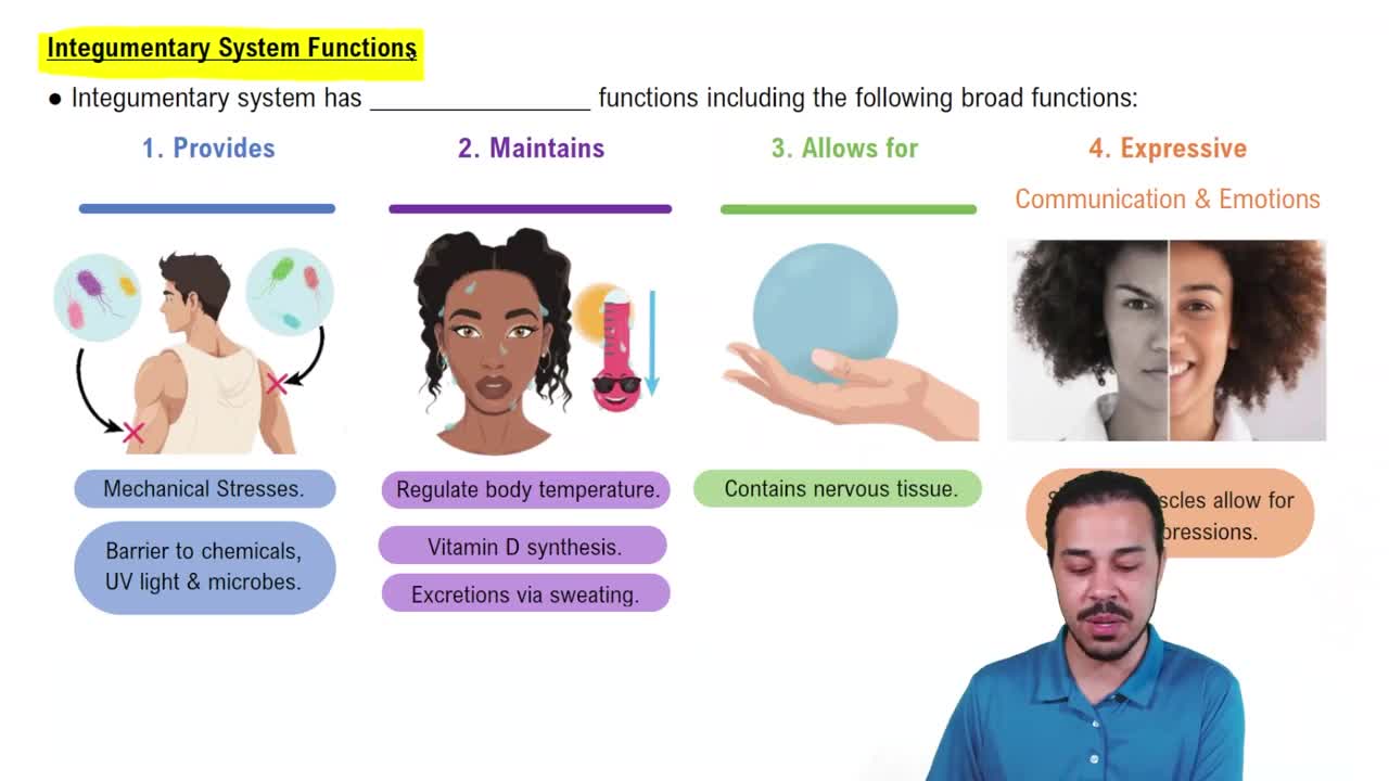
Terms in this set (40)
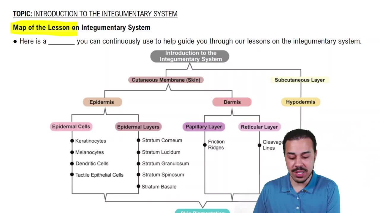
Keratinocytes are the primary cells in the epidermis responsible for producing keratin, a protein that strengthens the skin.
Merkel cells are sensory cells in the skin that detect touch stimuli.
Melanocytes produce melanin, the pigment that gives skin its color and protects against UV radiation.
Meissner’s corpuscles are touch receptors located in the dermal papillae, sensitive to light touch.
Dendritic cells are immune cells in the skin that detect pathogens and activate immune responses.
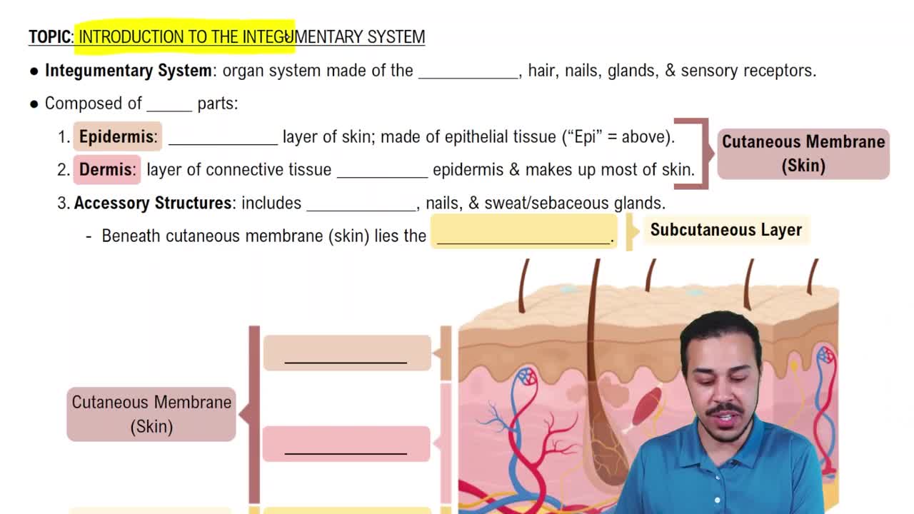
The stratum corneum is the outermost layer of the epidermis, composed of dead, cornified keratinocytes that provide a protective barrier.
The stratum granulosum contains keratinocytes that begin to die and accumulate keratohyalin granules, aiding in waterproofing.
The stratum spinosum is a layer of the epidermis where keratinocytes start producing keratin and are connected by desmosomes.
The stratum basale is the deepest epidermal layer where keratinocytes divide and new cells are generated.
Sudoriferous glands are sweat glands that help regulate body temperature through sweat secretion.
Sebaceous glands secrete sebum, an oily substance that lubricates and waterproofs the skin and hair.
Ceruminous glands are modified sweat glands in the ear canal that produce earwax (cerumen).
Multicellular exocrine glands are classified by their duct structure (simple or compound) and secretory unit shape (tubular, alveolar, or tubuloalveolar).
Jaundice is a yellowing of the skin and eyes caused by excess bilirubin in the blood.
Addison’s disease is a disorder causing insufficient adrenal hormones, leading to skin darkening and fatigue.
Cyanosis is a bluish discoloration of the skin due to low oxygen levels in the blood.
Dermal ridges are raised areas of the dermis that create fingerprints and improve grip.
The hypodermis is the subcutaneous layer beneath the dermis, composed mainly of fat and connective tissue for insulation and cushioning.
The arrector pili muscle is a small muscle attached to hair follicles that causes hair to stand up (goosebumps).
The dermis has two layers: the papillary layer (loose connective tissue) and the reticular layer (dense connective tissue).
Serous membranes line body cavities and secrete lubricating fluid to reduce friction between organs.
Mucous membranes line body cavities open to the exterior and secrete mucus for protection and lubrication.
Endothelium is a simple squamous epithelium lining blood vessels and lymphatic vessels.
Chondroblasts are cartilage-forming cells that produce the extracellular matrix of cartilage.
Elastic cartilage contains elastic fibers, providing flexibility; found in the ear and epiglottis.
Hyaline cartilage is the most common cartilage type, providing support with some flexibility; found in the nose and trachea.
Fibrocartilage is tough and dense, found in intervertebral discs and knee menisci for shock absorption.
Osseous tissue is bone tissue that provides structural support and protection.
Reticular tissue is a type of connective tissue with a network of reticular fibers supporting lymphoid organs.
Mesenchyme is embryonic connective tissue from which all connective tissues develop.
Collagen fibers provide tensile strength and structural support in connective tissues.
Microvilli increase surface area for absorption; found in organs like the small intestine and kidney tubules.
This epithelium appears layered but is a single layer; cilia move mucus; found in the respiratory tract.
Goblet cells secrete mucus to trap particles and lubricate surfaces, commonly found in respiratory and digestive tracts.
Simple squamous epithelium is a single layer of flat cells allowing rapid diffusion; found in lungs and blood vessels.
Stratified squamous epithelium has multiple layers for protection; found in skin, mouth, and esophagus.
The matrix consists of ground substance and fibers that provide support and determine tissue properties.
Connective tissue is made of cells, fibers (collagen, elastic, reticular), and ground substance.
Endocrine glands secrete hormones into the blood; exocrine glands release secretions through ducts to surfaces.
Muscular tissue enables movement; types include skeletal (voluntary), cardiac (heart), and smooth (involuntary) muscle.