 Back
Anatomy & Physiology Test 3 Study Guide
 04:06

Terms in this set (30)
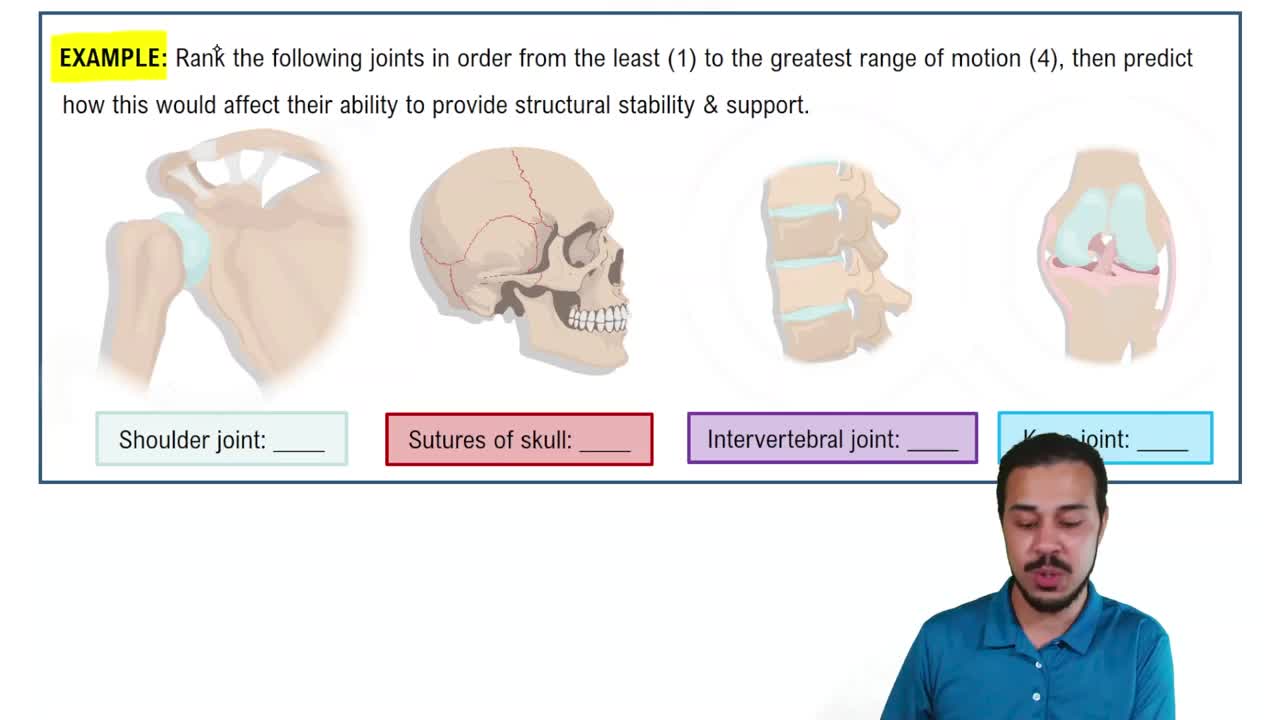
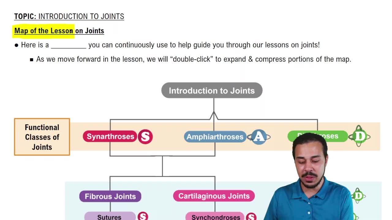
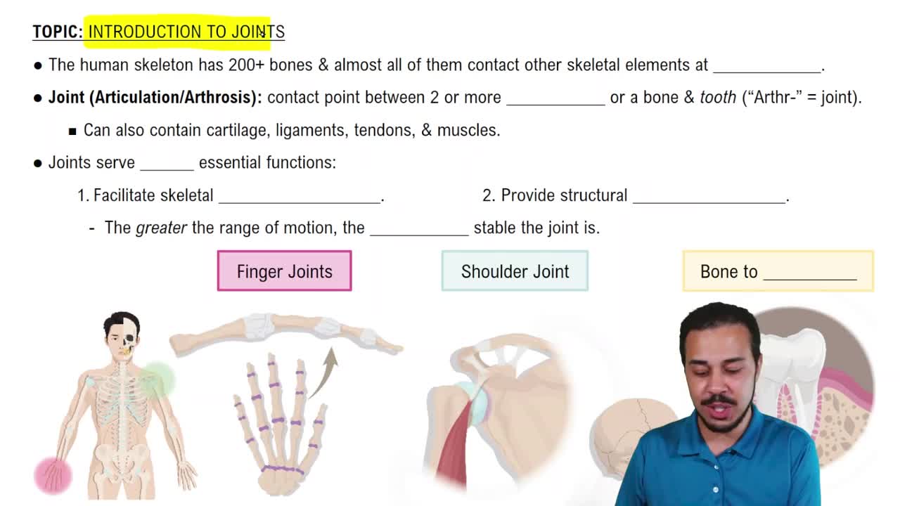
Synarthrosis is another name for an immovable joint, typically fibrous or bony fused joints like skull sutures or synostosis.
Suture, gomphosis, synchondrosis, and synostosis are the four types of immovable joints.
Movement away from the body's midline in the frontal plane, e.g., raising arms sideways from anatomical position.
Movement toward the body's midline in the frontal plane, e.g., lowering arms back to the sides.
Flexion, extension, abduction, adduction, opposition, reposition, and circumduction.
Movement of the thumb across the palm to touch the fingertips, combining abduction, flexion, and medial rotation.
Returning the thumb from opposition back to its anatomical position, involving extension, adduction, and lateral rotation.
Lateral ankle sprain involving the anterior talofibular ligament (ATFL), usually caused by foot inversion.
Movement of the foot upward toward the shin at the ankle joint.
Pointing the foot downward at the ankle joint, like pressing a gas pedal or standing on tiptoes.
The elbow is a synovial hinge joint allowing mainly flexion and extension.
Amphiarthrosis is a joint allowing slight movement.
Joints that move in three planes: flexion-extension, abduction-adduction, and rotation, e.g., ball-and-socket joints.
A biaxial synovial joint shaped like a saddle, found at the thumb carpometacarpal (CMC) joint.
Gliding, flexion, extension, rotation, abduction, adduction, circumduction, opposition, pronation, and supination depending on joint type.
Ball-and-socket joints, especially the shoulder, allow movement in all three planes.
Looking up at the ceiling by extending the neck backward beyond anatomical position.
The acromioclavicular ligament connects the clavicle to the acromion.
Glenohumeral ligaments, coracohumeral ligament, transverse humeral ligament, and coracoacromial ligament.
A condyloid (ellipsoid) synovial joint allowing biaxial movement: flexion/extension and abduction/adduction.
Diarthrosis is a freely movable joint, synonymous with synovial joints.
Weakening or tearing of the annulus fibrosus allows the nucleus pulposus to protrude, potentially compressing nerves.
Acetabular labrum, deep acetabulum, joint capsule, ligaments (iliofemoral, pubofemoral, ischiofemoral), and surrounding muscles.
Iliofemoral, pubofemoral, ischiofemoral ligaments, and ligamentum teres.
Shock absorption, load distribution, improved joint stability, and congruency in joints like the knee.
The patellar ligament supports the anterior knee, continuing from the quadriceps tendon to the tibial tuberosity.
The oblique popliteal ligament strengthens the posterior knee capsule.
The tibial collateral ligament (medial collateral ligament, MCL) resists valgus stress on the knee.
ACL prevents anterior tibial displacement; PCL prevents posterior tibial displacement relative to the femur.
The ankle (talocrural) joint is a synovial hinge joint allowing dorsiflexion and plantar flexion.