 Back
Anatomy & Physiology: The Heart
 6:13

Terms in this set (23)
The heart acts as a muscular double pump: the right side pumps oxygen-poor blood to the lungs (pulmonary circuit), and the left side pumps oxygen-rich blood to the body (systemic circuit).
The heart is located in the mediastinum, between the lungs, with the apex pointing to the left of the midline and the base as the broad posterior surface.
The pericardium has two primary layers: the fibrous pericardium (dense connective tissue) and the serous pericardium, which has a parietal and visceral layer (epicardium).
The heart wall consists of the epicardium (visceral serous pericardium), myocardium (cardiac muscle), and endocardium (endothelium lining internal walls).
The heart has two superior atria (right and left) that receive blood, and two inferior ventricles (right and left) that pump blood out.
Oxygen-poor blood enters the right atrium via the superior vena cava, inferior vena cava, and coronary sinus.
AV valves (tricuspid on right, bicuspid/mitral on left) prevent backflow of blood from ventricles to atria during ventricular contraction.
Chordae tendineae are tendon-like cords that anchor AV valve cusps to papillary muscles, preventing valve prolapse during ventricular contraction.
The aortic and pulmonary semilunar valves prevent blood from flowing back into the ventricles from the aorta and pulmonary trunk after contraction.
The cardiac skeleton is dense connective tissue surrounding valves; it anchors valve cusps, prevents overdilation, provides muscle insertion, and blocks direct electrical impulse spread.
Blood flows: right atrium → tricuspid valve → right ventricle → pulmonary semilunar valve → pulmonary trunk → lungs → pulmonary veins → left atrium → mitral valve → left ventricle → aortic semilunar valve → aorta → body.
The left ventricle has a wall three times thicker than the right ventricle because it pumps blood through the systemic circuit, which has higher resistance.
Cardiac muscle cells are short, branched, striated, with one or two nuclei, connected by intercalated discs containing gap junctions and desmosomes.
Intercalated discs electrically and mechanically connect cardiac muscle cells, allowing synchronized contraction through gap junctions and strong adhesion via desmosomes.
Calcium ions enter the sarcoplasm, triggering the sarcoplasmic reticulum to release more calcium, which initiates the sliding filament mechanism for contraction.
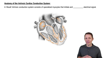
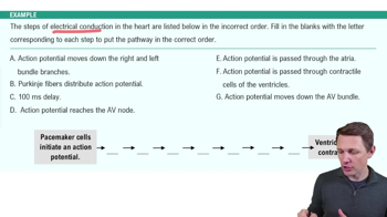
A network of specialized cardiac muscle cells that generate and conduct impulses, including the sinoatrial (SA) node, atrioventricular (AV) node, AV bundle, bundle branches, and Purkinje fibers.
The SA node acts as the heart's pacemaker, generating impulses that set the inherent rate of contraction.
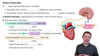
Parasympathetic fibers (vagus nerve) decrease heart rate; sympathetic fibers increase heart rate and contraction strength.
The right and left coronary arteries supply oxygenated blood to the myocardium, branching from the base of the aorta.
Coronary artery disease involves atherosclerosis causing reduced blood flow, leading to angina pectoris, myocardial infarction, or silent ischemia.
CHF is the progressive weakening of the heart, reducing its ability to meet oxygen demands, often causing heart enlargement and pulmonary hypertension.
Defects include ventricular septal defect, transposition of great vessels, coarctation of the aorta, and tetralogy of Fallot, affecting blood flow and oxygenation.
Aging causes valve thickening, reduced cardiac reserve, fibrosis of cardiac muscle, and decreased maximum heart rate, potentially leading to arrhythmias.