 Back
Anatomy & Physiology: The Skeletal System
 3:56

Terms in this set (28)
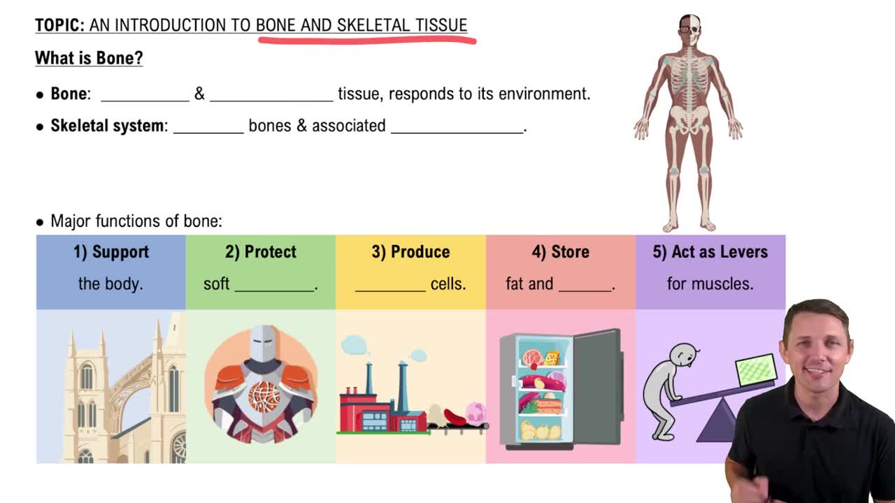
Bones, joints, cartilages, and ligaments.
Axial skeleton and appendicular skeleton.
Support the body, protect soft organs, allow movement, store minerals and fats, and blood cell formation (hematopoiesis).
Compact bone (dense and smooth) and spongy bone (needlelike pieces with many open spaces).
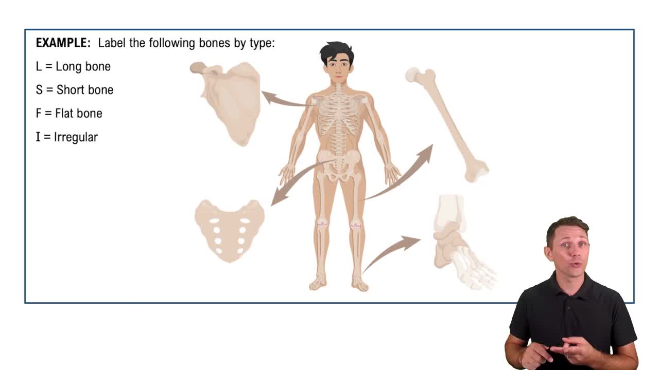
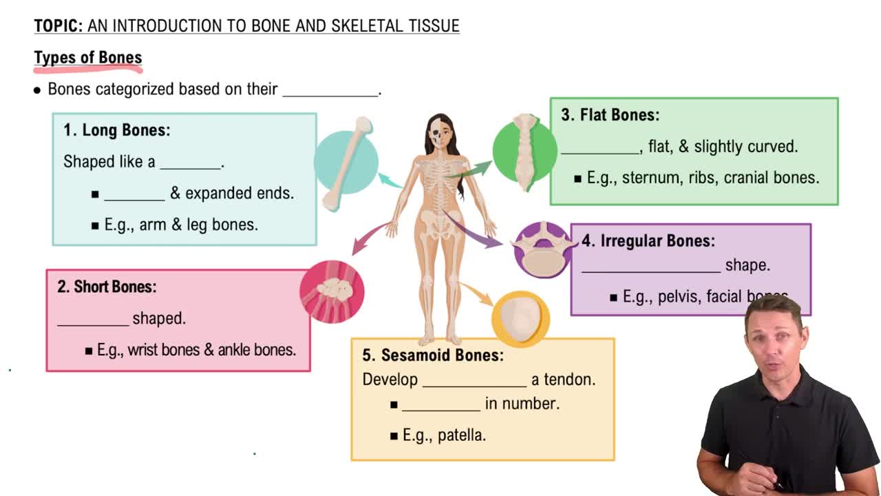
Long, flat, short, and irregular bones.
Longer than wide, shaft with enlarged ends, mostly compact bone with spongy bone at ends. Examples: femur, humerus.
Thin, flattened, usually curved, two layers of compact bone with spongy bone in between. Examples: skull bones, ribs, sternum.
Cube-shaped, mostly spongy bone with outer compact bone. Sesamoid bones form within tendons. Examples: carpals, tarsals, patella.
Irregular shape, do not fit other categories. Examples: vertebrae, hip bones.
Shaft that makes up most of the bone's length, composed of compact bone.
Fibrous connective tissue membrane covering the diaphysis, secured by perforating (Sharpey’s) fibers.
Ends of long bones, mostly spongy bone enclosed by thin compact bone layer, covered by articular cartilage.
Plate is hyaline cartilage causing lengthwise growth in young bones; line is its remnant in adults.
Contains yellow marrow (fat) in adults and red marrow (blood cell formation) in children.
Structural and functional unit of compact bone, consisting of central canal and concentric lamellae.
Tiny canals connecting osteocytes to the central canal, forming a nutrient transport system.
Organic collagen fibers provide flexibility and tensile strength; calcium salts provide hardness and resistance to compression.
Process of bone formation occurring on hyaline cartilage models or fibrous membranes.
Growth in bone width by osteoblasts adding matrix outside and osteoclasts removing bone inside the diaphysis.
Released when blood calcium is low; activates osteoclasts to break down bone and release calcium into blood.
Closed (simple): bone breaks but skin intact; open (compound): bone breaks and penetrates skin.
Hematoma formation, fibrocartilage callus formation, bony callus formation, and bone remodeling.
Skull, vertebral column, and bony thorax.
26 vertebrae: 7 cervical, 12 thoracic, 5 lumbar, plus sacrum and coccyx formed by fused vertebrae.
Primary curvatures (thoracic, sacral) present at birth; secondary curvatures (cervical, lumbar) develop after birth.
Plane, hinge, pivot, condylar, saddle, and ball-and-socket joints.
Articular cartilage reduces friction, articular capsule encloses joint, joint cavity contains synovial fluid, reinforcing ligaments stabilize joint.
Fibrous membranes in fetal skull allowing compression during birth and brain growth; ossify by about 2 years old.