 Back
BackAnatomy topic 1: Blood
 2:36
2:36
Terms in this set (115)
What is the pathway of blood through the cardiovascular system?
Heart → arteries → arterioles → capillaries → venules → veins → heart
What is plasma?
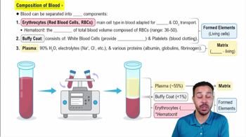
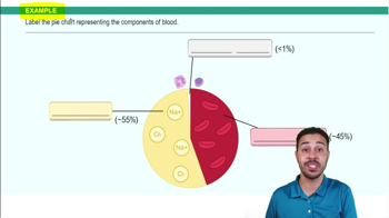
The nonliving fluid portion of blood that contains formed elements and dissolved substances.
What are the 3 formed elements of blood?
Erythrocytes, leukocytes, and platelets.
What color is arterial blood vs. venous blood?
Arterial blood = scarlet red. Venous blood = dark red.
What is the buffy coat?
A thin whitish layer between erythrocytes and plasma.
What is hematocrit?
The percentage of whole blood made up of red blood cells.
Normal adult hematocrit range?
Males: 47% ± 5%. Females: 42% ± 5%.
Normal blood pH?
7.35–7.45
What is albumin?
The most abundant plasma protein. Helps maintain osmotic pressure and transport substances.
What is gamma globulin?
The antibody portion of plasma.
Two functions of plasma electrolytes?
Buffering and maintaining osmotic pressure.
What organ produces clotting proteins?
Liver.
What are non-protein nitrogen substances?
Plasma urea, uric acid, creatinine, and ammonium salts.
What do alpha and beta globulins do?
Transport lipids, some metals, and fat-soluble vitamins.
What is unusual about mature erythrocytes?
They have no nucleus and essentially no organelles.
What proteins support the RBC membrane?
Actin and spectrin.
Average RBC count in adult men?
5.1–5.8 × 10⁶ RBC/mm³
Average RBC count in adult women?
4.3–5.2 × 10⁶ RBC/mm³
Average lifespan of an RBC?
100–120 days.
Average diameter of an RBC?
7.5 µm.
Most abundant protein in RBCs?
Hemoglobin.
How many heme groups are in one hemoglobin molecule?
4.
What is found in the center of each heme?
Iron ion, Fe²⁺.
How many O₂ molecules can one hemoglobin molecule carry?
4.
How many globin chains are in hemoglobin?
4 total: 2 alpha and 2 non-alpha chains.
Hematopoiesis vs. erythropoiesis?
Hematopoiesis = production of any formed blood element. Erythropoiesis = production of erythrocytes only.
Where does erythropoiesis occur in healthy adults?
Red bone marrow.
Order of erythropoiesis?
Hemocytoblast → proerythroblast → early erythroblast → late erythroblast → normoblast → reticulocyte → erythrocyte.
What nutrients are needed for erythropoiesis?
Nutrients, two B-complex vitamins, and iron.
What happens when blood O₂ decreases?
Kidney cells become hypoxic.
What hormone do the kidneys release during low O₂?
Erythropoietin.
What does erythropoietin do?
Stimulates red bone marrow to increase erythrocyte production.
What other organ secretes some erythropoietin?
Liver.
Ferritin and hemosiderin function?
Protein-iron complexes that store iron inside cells.
Transferrin function?
Binds and transports iron in the blood.
What cells destroy aged or damaged RBCs?
Macrophages.
Where are macrophages that destroy RBCs located?
Liver, spleen, and bone marrow.
What happens to hemoglobin when RBCs are destroyed?
It is broken into heme and globin.
What happens to globin?
It is broken into amino acids and reused.
What happens to heme?
Iron is removed and the rest is converted into bilirubin.
How does bilirubin leave the body?
Liver → bile → intestine → urobilin → eliminated in feces.
What is polycythemia?
Abnormally high erythrocyte production.
What is thalassemia?
Insufficient globin production.
What is aplastic anemia?
Reduced RBC production due to bone marrow destruction or inhibition.
What is hemolytic anemia?
Reduced RBC count due to premature or excessive RBC destruction
What is pernicious anemia?
Anemia caused by lack of vitamin B12.
What is hemorrhagic anemia?
Anemia caused by acute or chronic blood loss.
What is sickle cell anemia?
Anemia caused by a single amino acid substitution in the beta-globin chain.
What is iron deficiency anemia?
Lack of enough iron for hemoglobin synthesis.
How are leukocytes different from erythrocytes?
Leukocytes are larger and have lobed nuclei.
General function of leukocytes?
Protect the body through inflammatory and immune responses.
What is diapedesis?
Passage of blood cells through intact blood vessel walls into tissues.
What is amoeboid motion?
Movement using temporary cytoplasmic extensions.
What is positive chemotaxis?
Movement of a cell toward a chemical substance.
Which leukocytes are granulocytes?
Neutrophils, eosinophils, and basophils.
Which leukocytes are agranulocytes?
Lymphocytes and monocytes.
Leukocytes from most to least abundant?
Neutrophils → lymphocytes → monocytes → eosinophils → basophils.
Basophil function?
Synthesizes heparin and histamine.
Monocyte function?
Becomes a macrophage and increases in chronic infection.
Neutrophil function?
Phagocytosis, especially of bacteria; increases in acute infection.
Lymphocyte function?
Produces antibodies or directly fights antigens.
Eosinophil function?
Kills parasitic worms and helps control allergic/inflammatory reactions.
Granulocyte vs. agranulocyte lifespan?
Granulocytes live a few days. Agranulocytes may live from hours to years.
What cell produces platelets?
Megakaryocyte.
Normal adult platelet count?
150,000–400,000.
Main function of platelets?
Hemostasis.
Are platelets nucleated?
No.
Average platelet diameter?
2–4 µm.
What is leukocytosis?
Abnormally elevated leukocyte count.
What is leukopenia?
Abnormally low leukocyte count.
What causes infectious mononucleosis?
Epstein-Barr infection causing uncontrolled B-lymphocyte proliferation.
What is leukemia?
Malignant overproduction of leukocytes.
Acute myelocytic leukemia?
Abnormally increased production of monoblasts.
Chronic myelocytic leukemia?
Abnormally increased production of neutrophilic cells.
Chronic lymphocytic leukemia?
Abnormally increased production of lymphocytes.
Acute lymphocytic leukemia?
Abnormally increased production of lymphoblasts.
What is hemostasis?
The stoppage of bleeding.
What are the 3 mechanisms of hemostasis?
Vascular spasm, platelet plug formation, and coagulation.
What is vascular spasm?
Immediate constriction of a blood vessel after injury.
What happens when platelets contact an injured vessel wall?
They become activated, swell, form projections, and become sticky.
What does von Willebrand factor do?
Helps stabilize platelets by forming a bridge between collagen and platelets.
What chemicals strengthen vascular spasm?
Serotonin and thromboxane A₂.
What is a platelet plug?
A clump of platelets that plugs an opening in a vessel wall.
Platelet plugs work best for what type of wound?
Small tears.
What chemicals recruit more platelets?
ADP and thromboxane A₂.
Why is platelet plug formation positive feedback?
Activated platelets release chemicals that attract and activate more platelets.
Phase 1 of coagulation?
Formation of prothrombin activator through the intrinsic or extrinsic pathway.
Phase 2 of coagulation?
Conversion of prothrombin to thrombin.
Phase 3 of coagulation?
Conversion/polymerization of fibrinogen into fibrin.
Procoagulant vs. anticoagulant?
Procoagulants promote clotting. Anticoagulants inhibit clotting.
Intrinsic vs. extrinsic coagulation pathway?
Intrinsic factors are already in the blood. Extrinsic factors come from outside the blood.
Role of vitamin K in coagulation?
Required for synthesis of four clotting factors.
What is clot retraction?
Platelets contract and pull on fibrin strands, squeezing serum from the clot and pulling vessel edges together.
How are clots removed?
Fibrinolysis.
What happens during fibrinolysis?
Plasminogen is converted to plasmin, which helps dissolve the clot.
What is thrombocytopenia?
Platelet count below 50,000/mm³.
What is an embolus?
A free-floating intravascular clot.
What is a thrombus?
A clot attached to the unbroken wall of a blood vessel or heart chamber.
What is hemophilia?
A genetic defect in production of procoagulant factors.
What is disseminated intravascular coagulation (DIC)?
Widespread clotting that depletes clotting factors and can lead to inability to clot elsewhere.
What is an agglutinogen?
A genetically determined glycoprotein on RBCs that determines blood type.
What is an agglutinin?
Plasma proteins that combine with foreign agglutinogens and cause agglutination.
What antigens does A+ blood have?
A and Rh(D).
What antigens does B+ blood have?
B and Rh(D).
What antigens does O− blood have?
None.
What antigens does AB+ blood have?
A, B, and Rh(D).
What antibodies does type A blood produce?
Anti-B.
What antibodies does type B blood produce?
Anti-A.
What antibodies does type O blood produce?
Anti-A and anti-B.
What antibodies does type AB blood produce?
Neither anti-A nor anti-B.
Why is type O called the universal donor?
Type O RBCs do not contain A or B antigens.
Why is type AB called the universal recipient?
Type AB does not make anti-A or anti-B antibodies.
What causes erythroblastosis fetalis?
An Rh-negative mother carries an Rh-positive fetus and develops anti-D antibodies.
What do the mother's anti-D antibodies do?
They can attack the fetus's Rh-positive red blood cells.
How is erythroblastosis fetalis prevented?
Rh immune globulin prevents the mother from becoming sensitized to Rh-positive blood.