 Back
Cardiovascular System II: Blood Vessels - Anatomy & Physiology
 3:38

Terms in this set (32)
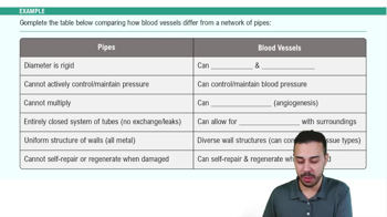
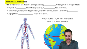
Transport blood to tissues for exchange of gases, nutrients, and wastes; regulate blood flow and pressure; secrete chemicals.
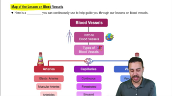
Arteries distribute blood away from the heart, capillaries enable exchange between blood and tissues, and veins collect blood and return it to the heart.
Innermost layer composed of endothelium continuous with heart lining; provides smooth surface to minimize friction; includes subendothelial connective tissue and internal elastic lamina.
Provide distensibility (stretch under pressure) and elasticity (recoil to original size after stretching).
Middle layer of smooth muscle cells and external elastic lamina; controls vessel diameter via vasoconstriction and vasodilation, regulated by sympathetic vasomotor nerves.
Outermost layer of dense irregular collagenous connective tissue; supports vessel and prevents overstretching; contains vasa vasorum supplying outer vessel layers.
Arteries have thicker tunica media and more elastic laminae due to higher pressure; veins have larger lumens, thinner walls, fewer elastic fibers, and often contain valves.
Elastic arteries (largest, near heart), muscular arteries (medium, distribute blood to organs), and arterioles (smallest, regulate flow into capillaries).
Metarterioles feed capillary beds; precapillary sphincters regulate blood flow into true capillaries by contracting or relaxing.
Veins outnumber arteries, have larger lumens, thinner walls, and serve as blood reservoirs holding about 70% of blood volume; contain valves to prevent backflow.
Connections between blood vessels providing collateral pathways; include arterial, venous, and arteriovenous anastomoses.
Study of blood flow physiology, driven by pressure gradients created by the heart, with flow inversely related to resistance.
Blood flow is directly proportional to pressure gradient and inversely proportional to resistance.
Peripheral resistance, cardiac output, and blood volume.
Blood vessel radius (inverse relation), blood viscosity, and vessel length.
CO = stroke volume × heart rate.
Pressure gradient (ΔP) = CO × PR (peripheral resistance).
MAP ≈ diastolic pressure + 1/3 (systolic pressure – diastolic pressure).
Difference between systolic and diastolic pressures, typically about 40 mm Hg.
Venous valves prevent backflow, skeletal muscle pumps squeeze veins, and respiratory pump uses pressure changes during breathing to propel blood.
Sympathetic stimulation increases heart rate and vasoconstriction raising BP; parasympathetic stimulation decreases heart rate lowering BP.
Negative feedback loop detecting blood pressure changes via stretch receptors to adjust sympathetic and parasympathetic output.
Epinephrine, norepinephrine, thyroid hormone increase CO; angiotensin-II causes vasoconstriction; ANP causes mild vasodilation.
Urinary and endocrine systems regulate blood volume via hormones (ADH, aldosterone, ANP, angiotensin-II) affecting water and sodium retention or excretion.
Walls consist of a single layer of endothelial cells and basal lamina; very thin (~0.2 μm) to allow exchange.
Continuous (least leaky), fenestrated (pores for rapid exchange), and sinusoidal (most leaky, large gaps).
Diffusion through gaps and membranes, osmosis, and transcytosis for larger molecules.
Arterioles constrict when pressure rises to reduce flow and dilate when pressure falls to maintain constant tissue perfusion.
Low oxygen and high CO2/hydrogen ions cause arteriolar dilation; high oxygen and low CO2 cause constriction.
Hydrostatic pressure pushes water out (~35 mm Hg arteriole end, 15 mm Hg venule end); osmotic pressure (~22 mm Hg) pulls water in due to plasma proteins.
Arteriole end: NFP positive, water filtered out; venule end: NFP negative, water absorbed back into capillary.
Increased capillary hydrostatic pressure, decreased osmotic pressure, or lymphatic obstruction leading to excess interstitial fluid.