The cardiovascular system consists of the heart, blood vessels, and blood. The heart pumps blood, which carries oxygen and nutrients, through the vessels.
Location and basic shape of the heart
The heart is a cone-shaped organ located slightly left in the thoracic cavity, posterior to the sternum, resting on the diaphragm in the mediastinum.
Four chambers of the heart
The heart has four chambers: two superior atria (right and left) and two inferior ventricles (right and left).
Difference between atria and ventricles
Atria receive blood from veins and pump it into ventricles; ventricles pump blood into arteries to the pulmonary or systemic circuits.
Pulmonary vs systemic circuits
The pulmonary circuit carries deoxygenated blood from the right heart to the lungs and back oxygenated to the left heart. The systemic circuit carries oxygenated blood from the left heart to the body and returns deoxygenated blood to the right heart.
Why is the pulmonary circuit low pressure and systemic circuit high pressure?
The pulmonary circuit pumps blood only to the lungs (short distance), so it is low pressure. The systemic circuit pumps blood to the entire body, requiring higher pressure.
Function of atrial natriuretic peptide (ANP)
ANP is a hormone produced by the atria that lowers blood pressure by decreasing sodium retention in kidneys, reducing blood volume and pressure.
Layers of the pericardium
The pericardium has two main layers: the tough outer fibrous pericardium and the inner serous pericardium, which includes the parietal and visceral layers separated by pericardial fluid.
Three layers of the heart wall
From superficial to deep: epicardium (visceral pericardium), myocardium (cardiac muscle and fibrous skeleton), and endocardium (endothelium lining the chambers).
Role of the fibrous skeleton of the heart
Provides structural support, anchors cardiac muscle cells, and electrically insulates atria from ventricles.
Great vessels of the heart
Include the superior and inferior vena cavae, pulmonary trunk, pulmonary veins, and aorta; they bring blood to and from the heart.
Difference between atrioventricular (AV) valves and semilunar valves
AV valves (tricuspid and mitral) prevent backflow from ventricles to atria; semilunar valves (pulmonary and aortic) prevent backflow from arteries to ventricles.
Function of chordae tendineae and papillary muscles
Chordae tendineae attach AV valve cusps to papillary muscles, which contract to prevent valve prolapse during ventricular contraction.
Coronary circulation purpose
Supplies oxygen and nutrients to the myocardium via coronary arteries and removes waste via coronary veins.
Common causes and consequences of coronary artery disease (CAD)
CAD is caused by plaque buildup reducing blood flow, leading to myocardial ischemia and potentially myocardial infarction (heart attack).
Autorhythmicity of cardiac muscle
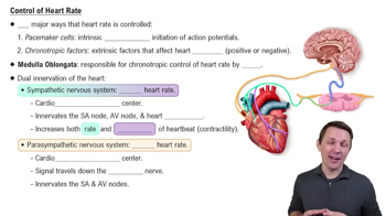
Cardiac muscle cells can generate their own action potentials without nervous system input, due to pacemaker cells.
Unique features of cardiac muscle cells
Branched cells with single nucleus, striated, abundant mitochondria, and intercalated discs containing desmosomes and gap junctions.
Phases of pacemaker cell action potential
Slow depolarization via nonspecific cation channels, full depolarization by calcium influx, repolarization by potassium efflux, then minimum potential phase.
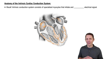
Components of the cardiac conduction system
Includes the sinoatrial (SA) node, atrioventricular (AV) node, AV bundle, bundle branches, and Purkinje fibers.
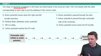
Sequence of electrical conduction in the heart
SA node → atrial conduction fibers → AV node (delay) → AV bundle → bundle branches → Purkinje fibers → ventricular contractile cells.
Phases of contractile cardiac muscle cell action potential
 Back
 6:13