 Back
Cardiovascular System: The Heart - Anatomy & Physiology
 6:13

Terms in this set (28)
The heart has two superior atria and two inferior ventricles.
The interatrial septum separates the atria, and the interventricular septum separates the ventricles.
The pericardium is a double-walled sac that protects the heart, anchors it to surrounding structures, and prevents overfilling.
Epicardium (visceral serous pericardium), myocardium (cardiac muscle layer), and endocardium (inner endothelial lining).
It anchors cardiac muscle fibers, supports heart valves and great vessels, and limits spread of action potentials to specific pathways.
Atrioventricular (AV) valves between atria and ventricles; Semilunar (SL) valves between ventricles and major arteries (aorta and pulmonary trunk).
Chordae tendineae anchor valve cusps to papillary muscles, holding them closed during ventricular contraction.
They prevent backflow from arteries into ventricles by opening when ventricular pressure rises and closing when it falls.
Each side of the heart pumps blood to its own circuit: the right side to the pulmonary circuit and the left side to the systemic circuit.
The right ventricle is thinner and crescent-shaped; the left ventricle is thicker and round-shaped, wrapping around the right ventricle.
They contain desmosomes and gap junctions that mechanically and electrically connect cardiac muscle cells, enabling synchronized contraction.
Cardiac muscle cells are self-excitable, contract as a unit, have longer action potentials with a plateau phase, and rely almost exclusively on aerobic respiration.
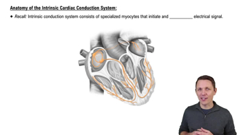
A network of autorhythmic cells that initiate and distribute impulses to coordinate heart depolarization and contraction.
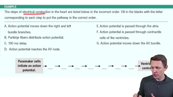
1. Sinoatrial (SA) node → 2. Atrioventricular (AV) node → 3. AV bundle (bundle of His) → 4. Right and left bundle branches → 5. Purkinje fibers (subendocardial conducting network).
The SA node is the heart's pacemaker, generating impulses about 75 times per minute to set the heart's rhythm.
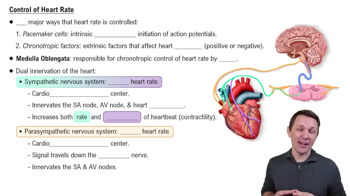
The sympathetic nervous system increases heart rate and contractility; the parasympathetic nervous system decreases heart rate via the vagus nerve.
Includes ventricular filling, isovolumic contraction, ventricular ejection, and isovolumic relaxation, coordinating blood flow through the heart.
First sound (lub) is closing of AV valves; second sound (dup) is closing of semilunar valves during the cardiac cycle.
Stroke volume (SV) is the volume of blood pumped per beat; cardiac output (CO) is SV multiplied by heart rate (HR), representing blood pumped per minute.
Preload (degree of muscle stretch), contractility (strength of contraction), and afterload (arterial pressure opposing ejection).
Increased preload (stretch) leads to increased stroke volume due to optimal overlap of cardiac muscle fibers.
Increased contractility lowers end systolic volume and increases stroke volume by enhancing calcium availability for contraction.
Afterload is the pressure ventricles must overcome to eject blood; increased afterload (e.g., hypertension) raises end systolic volume and reduces stroke volume.
Positive chronotropic factors increase heart rate; negative chronotropic factors decrease heart rate.
Epinephrine and norepinephrine increase heart rate and contractility; thyroxine increases heart rate and enhances effects of catecholamines.
Tachycardia: abnormally fast heart rate; Bradycardia: abnormally slow heart rate, which can be normal in athletes or pathological.
A condition where cardiac output is insufficient to meet tissue needs, caused by weakened myocardium from factors like coronary atherosclerosis or high blood pressure.
Foramen ovale connects atria; ductus arteriosus connects pulmonary trunk to aorta; both close after birth.