 Back
Ch 6 Bones and Bone Tissue
 3:56

Terms in this set (20)
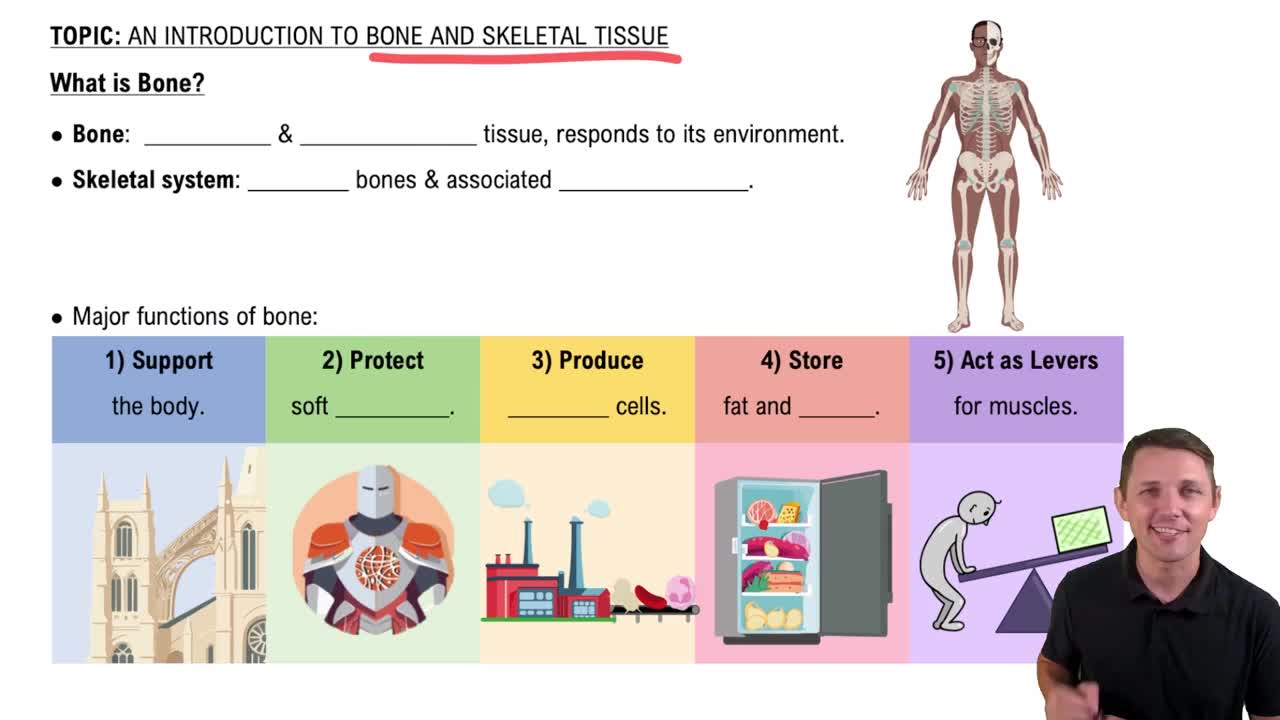
Protection, mineral storage (Ca, P, Mg), hematopoiesis (blood cell formation), movement, support, fat storage, and hormone production.
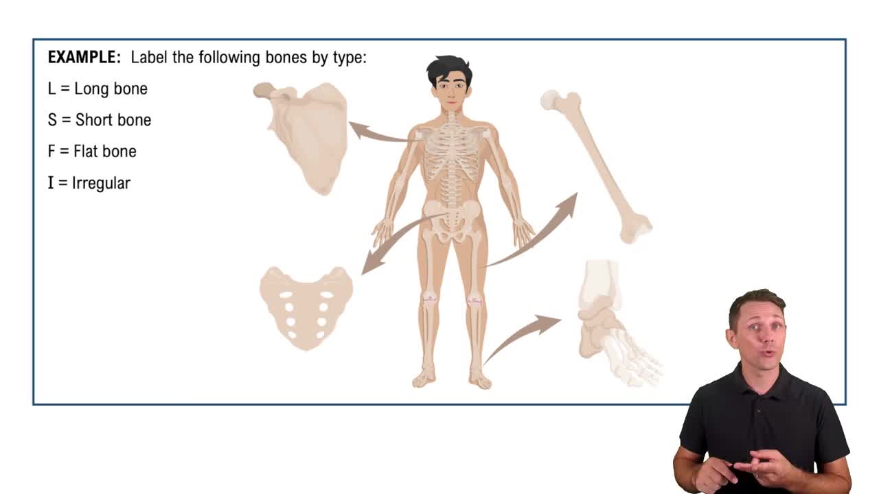
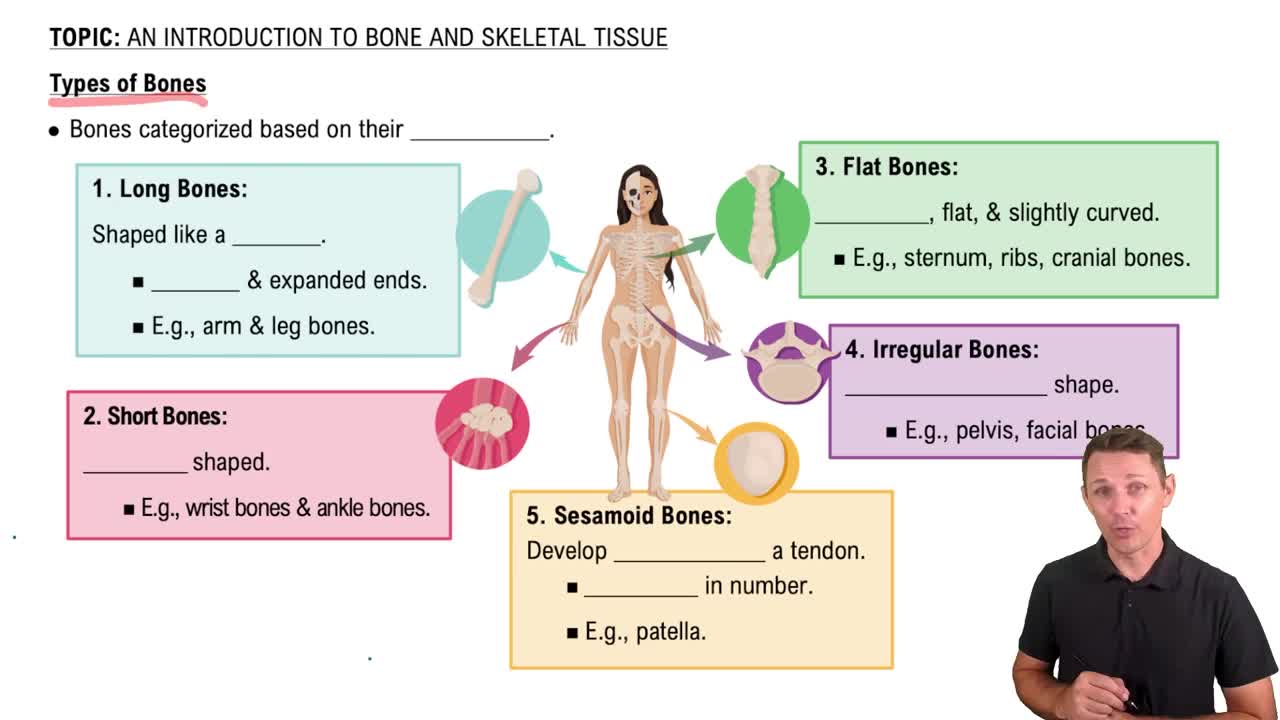
Long bones (longer than wide), short bones (about as long as wide), flat bones (broad, flat, thin), irregular bones (shape does not fit other classes), and sesamoid bones (round, flat bones found within tendons).
Includes epiphysis (ends), diaphysis (shaft), periosteum (outer fibrous and osteogenic layers), articular cartilage, nutrient foramina, endosteum, medullary cavity, compact bone, and spongy (cancellous) bone.
Outer dense irregular collagenous layer and inner osteogenic layer; collagen fibers extend into bone matrix as perforating fibers, providing attachment and bone growth/repair.
Red marrow: hematopoietic stem cell niche, highly vascularized. Yellow marrow: fat storage, also vascularized. Composition changes with age.
Inorganic matrix: mostly hydroxyapatite (calcium phosphate), provides compression resistance. Organic matrix (osteoid): mostly collagen and glycosaminoglycans (GAGs), provides tensile strength.
Removing organic matrix (collagen) makes bone brittle and prone to shattering. Removing inorganic matrix (minerals) makes bone unable to resist compression.
Rickets: calcium deficiency causing soft, deformed bones. Osteogenesis imperfecta: collagen defect causing brittle bones.
Mesenchymal stem cells in periosteum and endosteum that differentiate into osteoblasts; important for fracture repair.
Bone-forming cells found in inner periosteum and endosteum; secrete collagen, osteocalcin, calcium, and phosphate to form osteoid that mineralizes into bone.
Mature osteoblasts trapped in lacunae within bone matrix; over 90% of bone cells; have dendritic processes in canaliculi for sensing strain and cell communication.
Multinucleated cells from bone marrow that resorb bone by secreting H+ ions and enzymes; release minerals into bloodstream.
Organized into osteons (Haversian systems) with concentric lamellae around central canals; osteocytes in lacunae connected by canaliculi; perforating (Volkmann) canals connect osteons.
Rings of collagen fibers in bone matrix; collagen fibers alternate orientation between lamellae to increase strength.
Composed of trabeculae (rod or plate-like structures) with concentric lamellae but no osteons or canals; helps distribute stress efficiently.
Bone formation within mesenchymal membrane; mesenchymal stem cells differentiate into osteoblasts forming osteoid; forms skull and clavicles.
Bone formation within hyaline cartilage scaffold; chondrocytes die, cartilage calcifies, osteoblasts form bone collar and spongy bone; forms most other bones.
Longitudinal growth: lengthening at epiphyseal plate, stops after adolescence. Appositional growth: width growth by bone deposition under periosteum, continues throughout life.
Bone architecture adapts to mechanical stresses placed on it; bone mass and density increase in response to greater load.
Continuous process replacing about 10% of bone per year; balances bone resorption by osteoclasts and formation by osteoblasts to maintain strength and mineral homeostasis.