 Back
Backchapter 1
 3:06
3:06
Terms in this set (73)
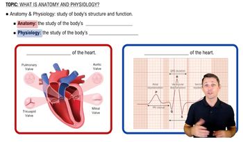
Anatomy
the study of internal and external body structures and their physical relationships among other body parts
Physiology
the study of how living organisms perform their vital functions
Anatomy and physiology are closely integrated
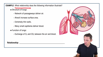
– The principle of complementarity of structure and
function states that all specific functions are
performed by specific structures, and the form of a
structure relates to its function.
Human anatomy
study of the structure of the human body
Gross anatomy
or macroscopic anatomy, examines large structures, visible with the naked eye
Microscopic anatomy
examines structures that can only be seen using magnification, such as cells and
molecules
Surface anatomy
anatomy of body surface
Regional anatomy
anatomy of specific body areas
Sectional anatomy
understanding the relationship of body structures by examining cross sections
Systemic anatomy
anatomy of organ systems
Clinical anatomy
anatomy as used in the clinical practice
Pathological anatomy
anatomical changes during illness
Radiologic anatomy
anatomical structures seen usingimaging techniques
Surgical anatomy
anatomical landmarks important in surgery
Developmental anatomy
anatomical changes from fertilization toadulthood
Embryology
study of early developmental processes
Cytology
study of the structure of cells
Histology
study of the structure of tissues
Human physiology
the study of the function of the human body
Cell physiology
study of the function of cells and their chemical processes
Organ physiology
study of the function of specific organs
Systemic physiology
study of function of organ systems
Pathological physiology
study of effects of diseases on organs or systems
Signs
objective disease indications (such as a fever)
Symptoms
subjective disease indication (such as tiredness)
Learning Outcome
Identify the major levels of organization in organisms from the simplest to the most complex and identify major components of each organ system.
Atoms
are the smallest stable units of matter
Molecules
consist of two or more atoms
Cells
the smallest living units in the body
Tissue level
A tissue is a group of cells working together to perform specific functions
Organ level- Organs
are made of two or more tissues working together to perform specific functions
Organ system level▪ An organ system
is a group of organs interacting for a particular function
Organism level- organism
An organism is an individual life form
Learning Outcome
Describe the origins of anatomical and physiological terms and explain the significance of standardizing terms.
Medical terminology
Involves using word roots, prefixes, suffixes and combining forms to build terms related to the body in health and disease
Learning Outcome
Use anatomical terms to describe body regions, body sections, and relative positions.
Surface anatomy
locating structures on or near the body surface
Anatomical position
standard anatomical reference position with hands at the sides, palms facing forward and feet together
Anterior view
body in anatomical position from the front
Posterior view
body in anatomical position from the back
Supine
body lying face up
Prone
body lying face down
Sectional anatomy
A section is a slice through a three-dimensional object
▪ Used to visualize internal organization
▪ Important for interpreting medical scans
Sectional plane
a single view or slice along a two-dimensional flat surface
Frontal (coronal) plane
a vertical plane that divides the body or organ into anterior and posterior portions
Sectional plane (cont.)– Sagittal plane
a vertical plane that divides the body or organ into left and right portions
Midsagittal
plane lies in the middle
Parasagittal
plane is offset from the middle
Transverse (horizontal) plane
a horizontal plane that divides the body or organ into superior and inferior portions transverse section (cross section)
Body cavities
– closed, fluid filled cavities that are lined by a thin serous membrane; contain the vital organs(viscera) of the trunk
Functions of body cavities
1. Protect delicate organs from shocks and impacts
2. Permit significant changes in the size and shape of internal organs
Serous membrane (serosa)
– Lines body cavities and covers organs
– Consists of parietal and visceral layers
▪ Parietal serosa lines cavity
▪ Visceral serosa covers organ
– Serous fluid moistens membranes and reduces friction
Thoracic cavity
– Deep to the chest wall of the thoracic region
– Divided by from the abdominopelvic cavity by the
diaphragm
The thoracic cavity contains:
▪ Right and left pleural cavities which surround the
lungs
Pericardial
cavity which surrounds the heart
Mediastinum
a mass of connective tissue that
stabilizes the trachea, esophagus, thymus, and
largest vessels of the heart; also contains the
pericardial cavity
Abdominopelvic cavity
Abdominopelvic cavity
– Deep to the abdominal and pelvic walls
– Extends from the diaphragm to the pelvis
– The abdominopelvic cavity contains:
▪ Superior abdominal cavity
▪ Inferior pelvic cavity
Peritoneal cavity
space within the
abdominopelvic cavity lined with peritoneum
– Parietal peritoneum lines the internal body wall
– Visceral peritoneum covers the organs
Abdominal cavity
– Extends from the diaphragm to the top of the pelvic
bones
– Contains digestive organs
– Most abdominal organs are enclosed by the peritoneal
cavity
Retroperitoneal space
▪ Area between the parietal peritoneum and the back
of the muscular body wall
▪ Contains organs such as the pancreas and kidneys
Pelvic cavity
– Medial to the pelvic bones
– Contains the reproductive organs, rectum, and urinary
bladder
– Contains the inferior portion of the peritoneal cavity
Infraperitoneal
organs which extend inferior to the
peritoneal cavity, such as the urinary bladder, distal
portion of ureters, and large intestine
Homeostasis
-the continuous physiological processes
that establish a relatively stable internal environment.
– Physiological processes and systems respond to
external and internal changes to keep variables within
normal ranges (body temperature, blood pressure, etcetera)
Homeostatic regulation
– the adjustment of
physiological systems to preserve homeostasis
– Mechanisms of homeostatic regulatio
Autoregulation
automatic, local response to an
environmental change in a cell, tissue, or organ
Extrinsic regulation
– responses of organ
systems controlled by the nervous system (through
electrical signals) or the endocrine system
(through chemical messengers)
– Nervous system directs rapid, short-term
responses
– Endocrine system directs slower, more long-
term responses
A homeostatic regulatory mechanism consists of
1. Receptor – a sensor that detects the stimulus or
change
2. Control center – receives and processes the
information and sends out commands
3. Effector – a cell of organ that carries out the
commands of the control center
Negative feedback
- type of regulation that opposes
variation from normal
– The response of the effector negates the original
stimulus
– Helps maintain variables within a normal range
Positive feedback
– type of regulation that enhances
variation from normal
– The initial stimulus produces a response that amplifies
the original change in conditions
Positive feedback loops
are used when a potentially
dangerous process must be completed quickly to
restore homeostasis
Systems integration
– systems work together to maintain
homeostasis
– Any adjustments made by one physiological system have
direct and indirect effects on a variety of other systems
Homeostasis is a state of equilibrium
Opposing processes or forces are in balance
Dynamic equilibrium
physiological systems are
continually adapting and adjusting to changing conditions.
The normal range of values can therefore vary depending
on conditions
Failure to maintain homeostasis leads to disease and possibly
death