 Back
Chapter 6 Bone Tissue Review - Anatomy & Physiology
 2:05

Terms in this set (28)
Long bones are longer than wide; short bones are cube-shaped; flat bones are thin and flat; irregular bones have complex shapes; sesamoid bones develop within tendons.
Intramembranous bone forms directly from mesenchymal tissue; endochondral bone forms by replacing hyaline cartilage.
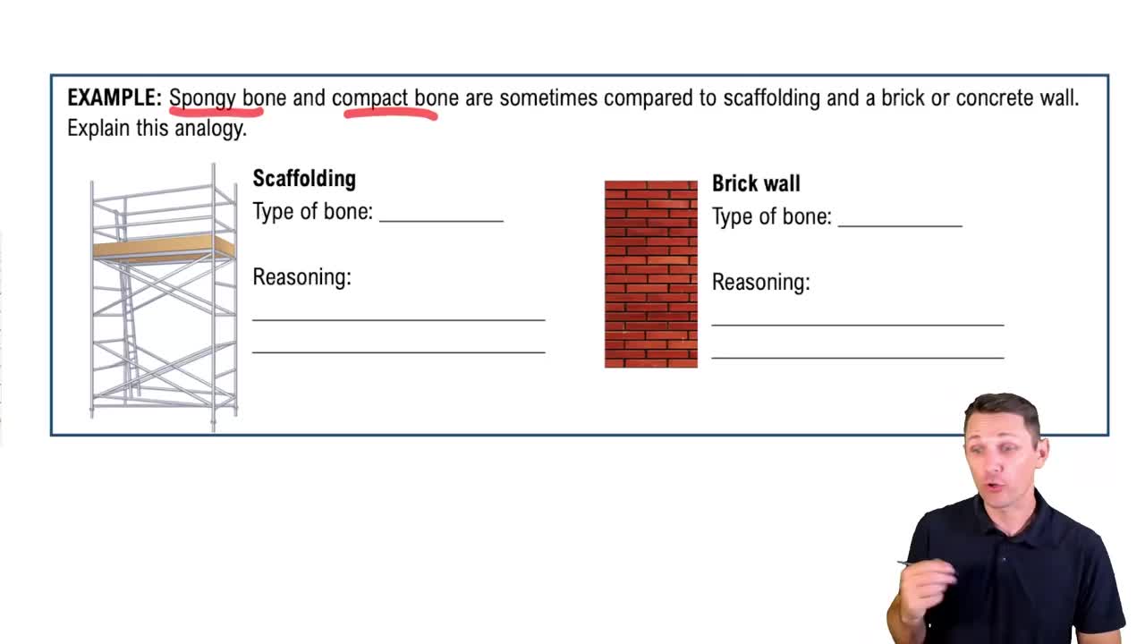
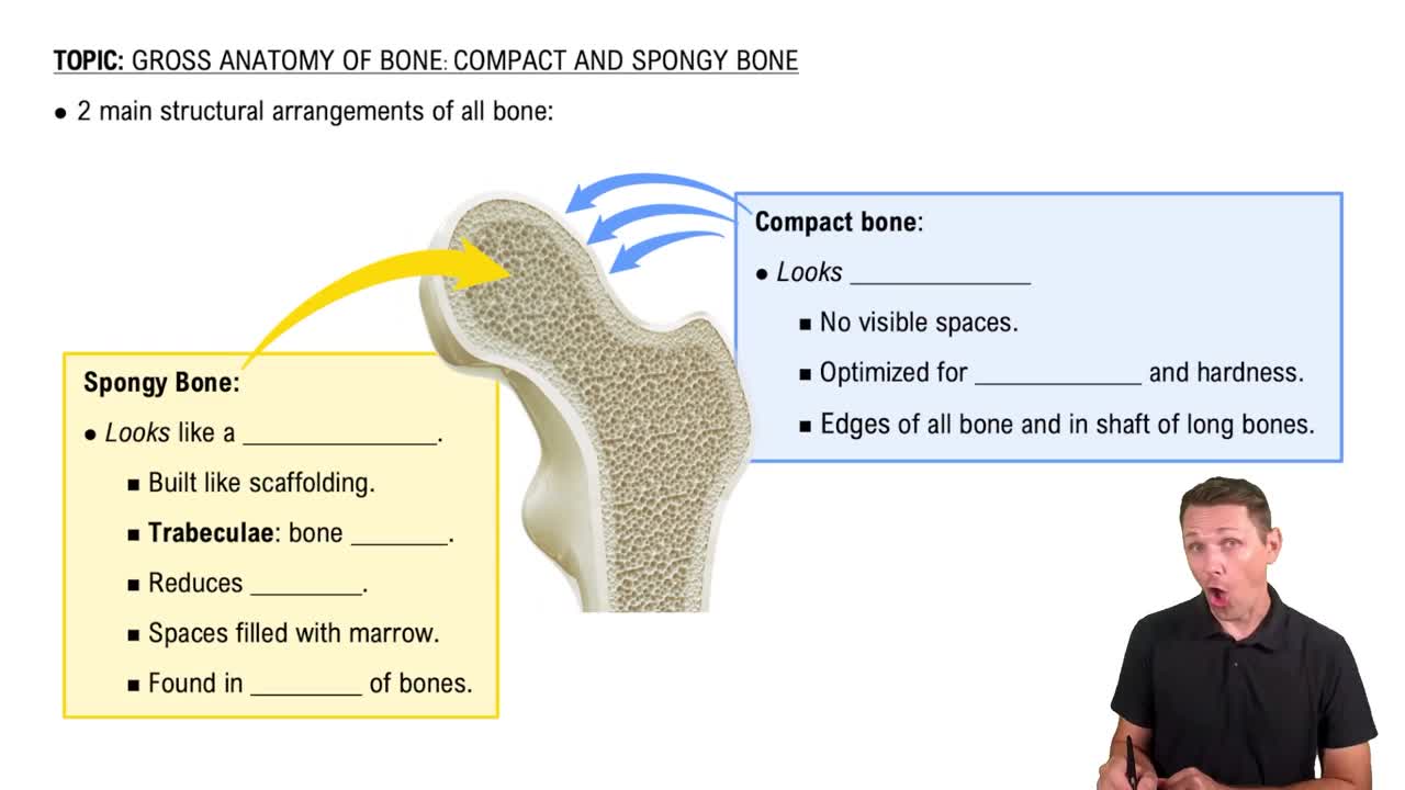
Compact bone is dense and forms the outer layer; spongy bone is porous, found inside bones, and contains trabeculae.
A dense connective tissue membrane covering the outer surface of bones, containing osteoblasts for growth and repair.
A thin membrane lining the marrow cavity and inner surfaces of bones, containing osteogenic cells.
The expanded end of a long bone, usually covered with articular cartilage.
The shaft or central part of a long bone.
A mature bone cell that maintains the bone matrix and resides in lacunae.
A bone-forming cell that secretes osteoid and initiates calcification.
A large cell that resorbs or breaks down bone matrix during remodeling.
The structural unit of compact bone, consisting of concentric lamellae around a central canal containing blood vessels and nerves.
Concentric rings of calcified matrix in compact bone surrounding the central canal.
Microscopic canals connecting lacunae, allowing osteocytes to communicate and exchange nutrients.
The central cavity within the diaphysis that contains bone marrow.
A hyaline cartilage plate in the metaphysis where bone growth occurs in children and adolescents.
Mesenchymal cells differentiate into osteoblasts, secrete osteoid, which calcifies, forming spongy bone that remodels into compact bone.
Hyaline cartilage model forms, cartilage calcifies, chondrocytes die, and bone tissue replaces cartilage from the diaphysis outward.
Support, protection, movement, mineral storage, and blood cell production.
Balance between osteoblast bone formation and osteoclast bone resorption to maintain bone strength and calcium levels.
Requires adequate nutrition, hormones (growth hormone, thyroid hormone), mechanical stress, and vitamin D.
Hematoma formation, fibrocartilaginous callus formation, bony callus formation, and bone remodeling.
PTH increases blood calcium by stimulating osteoclasts, increasing intestinal absorption, and reducing kidney excretion.
Calcitonin lowers blood calcium by inhibiting osteoclast activity and promoting calcium deposition in bone.
Process is a projection; tubercle is a small rounded projection; trochanter is a large, blunt projection.
Foramen is a hole for blood vessels/nerves; fissure is a narrow slit; sinus is a cavity within a bone.
The region of a long bone between the diaphysis and epiphysis containing the epiphyseal plate.
Consists of trabeculae with spaces filled by marrow; lacks osteons found in compact bone.
Organized into osteons with central canals, lamellae, lacunae, and canaliculi.