Skip to main content
Anatomy & Physiology
My Course
Learn
Exam Prep
AI Tutor
Study Guides
Textbook Solutions
Flashcards
Explore
Try the app
My Course
Learn
Exam Prep
AI Tutor
Study Guides
Textbook Solutions
Flashcards
Explore
Try the app
Back
Heart Anatomy and Major Vessels
You can tap to flip the card.
Aorta
You can tap to flip the card.
👆
Aorta
The largest artery in the body that carries oxygenated blood from the left ventricle to the systemic circulation.
Track progress
Control buttons has been changed to "navigation" mode.
1/20
Recommended videos
Guided course
6:13
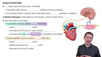
Control of Heart Rate
8267
views
172
rank
1
comments
Guided course
8:41
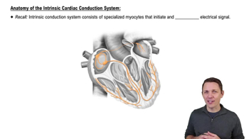
Anatomy of the Intrinsic Cardiac Conduction System
14196
views
340
rank
3
comments
Guided course
3:42
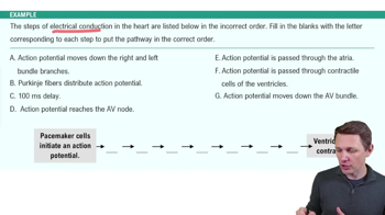
Electrical Conduction System of the Heart Example 2
7742
views
190
rank
Terms in this set (20)
Hide definitions
Aorta
The largest artery in the body that carries oxygenated blood from the left ventricle to the systemic circulation.
Brachiocephalic Trunk
A major artery branching from the aortic arch that supplies blood to the right arm and the head and neck.
Left Common Carotid Artery
An artery arising from the aortic arch that supplies oxygenated blood to the left side of the head and neck.
Left Subclavian Artery
An artery branching from the aortic arch that supplies blood to the left arm.
Superior & Inferior Venae Cavae
Large veins that return deoxygenated blood from the body to the right atrium; superior from upper body, inferior from lower body.
Pulmonary Trunk
A vessel that carries deoxygenated blood from the right ventricle to the lungs for oxygenation.
Pulmonary Veins
Vessels that carry oxygenated blood from the lungs to the left atrium.
Right & Left Atria and Auricles
The upper chambers of the heart that receive blood; auricles are small appendages that increase atrial volume.
Right & Left Ventricles
The lower chambers of the heart responsible for pumping blood out to the lungs and body.
Apex & Base of the Heart
The apex is the pointed tip of the heart directed downward; the base is the broad top where major vessels attach.
Interventricular Septum
The muscular wall separating the right and left ventricles.
Moderator Band
A muscular band in the right ventricle that carries part of the conduction system and prevents overexpansion.
Tricuspid Valve
The valve between the right atrium and right ventricle that prevents backflow of blood.
Mitral (Bicuspid) Valve
The valve between the left atrium and left ventricle that prevents backflow of blood.
Pulmonic Semilunar Valve
The valve between the right ventricle and pulmonary trunk that prevents backflow into the ventricle.
Aortic Semilunar Valve
The valve between the left ventricle and aorta that prevents backflow into the ventricle.
Chordae Tendineae
Tendinous cords that connect papillary muscles to the atrioventricular valves to prevent valve prolapse.
Papillary Muscles
Muscles located in the ventricles that anchor chordae tendineae and contract to prevent valve inversion.
Epicardium
The outer layer of the heart wall, also known as the visceral layer of the pericardium.
Pericardium
A double-walled sac surrounding the heart that provides protection and reduces friction during heartbeats.
 BackBack
 6:13