 Back
Heart Anatomy and Physiology
 6:13

Terms in this set (20)
The heart is roughly the size of a fist, conical in shape, located in the thorax between the lungs, slightly left of the midline, and oriented with the apex pointing down and to the left.
The heart is covered by the pericardium, which includes the fibrous pericardium and the serous pericardium (parietal and visceral layers).
Epicardium: outer protective layer; Myocardium: thick muscular middle layer responsible for contraction; Endocardium: inner lining that contacts blood.
Right atrium (receives deoxygenated blood via superior/inferior vena cava), right ventricle (pumps to lungs via pulmonary trunk), left atrium (receives oxygenated blood via pulmonary veins), left ventricle (pumps to body via aorta).
Atrioventricular valves (tricuspid and mitral) prevent backflow into atria; Semilunar valves (pulmonary and aortic) prevent backflow from arteries into ventricles.
Valves open and close in response to pressure changes during the cardiac cycle, ensuring unidirectional blood flow.
Blood flows: body → right atrium → tricuspid valve → right ventricle → pulmonary valve → lungs → left atrium → mitral valve → left ventricle → aortic valve → body.
Left coronary artery branches into the anterior interventricular and circumflex arteries; right coronary artery branches into the marginal and posterior interventricular arteries.
Cardiac muscle is striated, involuntary, branched, with intercalated discs for electrical coupling, allowing synchronized contraction unlike skeletal muscle.
Action potential triggers Ca2+ influx, which induces Ca2+ release from the sarcoplasmic reticulum, leading to muscle contraction.
Pacemaker cells have spontaneous depolarization and no stable resting potential; contractile cells have a stable resting potential and a plateau phase for sustained contraction.
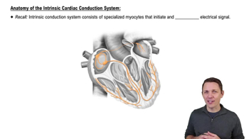
Includes the sinoatrial (SA) node, atrioventricular (AV) node, bundle of His, bundle branches, and Purkinje fibers.
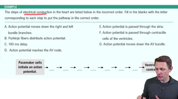
Impulse starts at SA node → AV node → bundle of His → bundle branches → Purkinje fibers → ventricular contraction.
P wave: atrial depolarization; QRS complex: ventricular depolarization; T wave: ventricular repolarization; intervals represent conduction times.
Includes arrhythmias, heart block, ischemia, and myocardial infarction indicated by changes in wave shape, timing, or intervals.
Includes atrial systole, ventricular systole, and diastole phases coordinating contraction and relaxation for blood flow.
Normal sounds: lub-dub from valve closures; murmurs are abnormal sounds from turbulent blood flow due to valve issues.
Include preload (venous return), contractility (strength of contraction), and afterload (resistance to ejection).
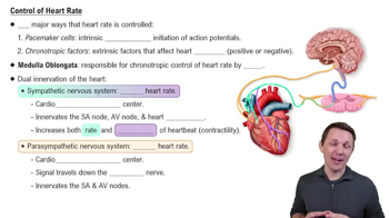
Include autonomic nervous system input, hormones, body temperature, and fitness level.
Sympathetic stimulation increases heart rate and contractility; parasympathetic stimulation decreases heart rate, modulating cardiac output.