Skip to main content
Anatomy & Physiology
My Course
Learn
Exam Prep
AI Tutor
Study Guides
Textbook Solutions
Flashcards
Explore
Try the app
My Course
Learn
Exam Prep
AI Tutor
Study Guides
Textbook Solutions
Flashcards
Explore
Try the app
Back
Heart Physiology and Cardiac Function
You can tap to flip the card.
What does the T wave represent on an ECG?
You can tap to flip the card.
👆
What does the T wave represent on an ECG?
The
T wave
represents
ventricular repolarization
.
Track progress
Control buttons has been changed to "navigation" mode.
1/27
Recommended videos
Guided course
6:13
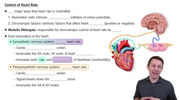
Control of Heart Rate
8267
views
172
rank
1
comments
Guided course
8:41
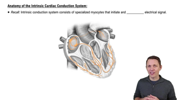
Anatomy of the Intrinsic Cardiac Conduction System
14197
views
340
rank
3
comments
Guided course
3:42
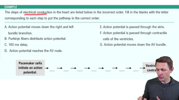
Electrical Conduction System of the Heart Example 2
7742
views
190
rank
Terms in this set (27)
Hide definitions
What does the T wave represent on an ECG?
The
T wave
represents
ventricular repolarization
.
What does the QRS complex represent on an ECG?
The
QRS complex
represents
ventricular depolarization
.
What does the P wave represent on an ECG?
The
P wave
represents
atrial depolarization
.
What is a ventricular septal defect?
A
ventricular septal defect
is a hole in the wall between the two ventricles.
What is an atrial septal defect?
An
atrial septal defect
is a defect in the wall between the two atria.
Which vessels return oxygenated blood from the lungs to the heart?
The
pulmonary veins
return oxygenated blood from the lungs to the heart.
Which ventricle pumps blood into the pulmonary circuit?
The
right ventricle
pumps blood into the pulmonary circuit.
What are the external flaps on the outside of the atria called?
The
auricles
are the external flaps on the outside of the atria that allow some extended volume.
Which channels are responsible for the plateau phase in cardiac action potentials?
Calcium
channels are responsible for the plateau phase.
What is the name of the vein formed by the veins of the heart muscle that empties into the right atrium?
The veins merge to form the
coronary sinus
which empties into the
right atrium
.
What is the fetal lung bypass from the pulmonary trunk to the aorta called, and what does it become after birth?
The fetal lung bypass is the
ductus arteriosus
, which closes at birth and becomes the
ligamentum arteriosum
.
What are the stabilizing strings of the tricuspid and bicuspid valves called?
They are called
chordae tendineae
.
What are the muscular ridges inside the ventricles called?
The muscular ridges inside the ventricles are called
trabeculae carneae
.
Are the AV valves open or closed during ventricular systole?
During ventricular systole, the
AV valves are closed
.
Are the semilunar valves open or closed during ventricular systole (ventricular ejection phase)?
During ventricular systole, the
semilunar valves are open
.
Are the semilunar and AV valves open or closed during isovolumetric relaxation?
During isovolumetric relaxation, both the
semilunar valves and AV valves are closed
.
Where does the heartbeat originate and what is its inherent rate?
The heartbeat originates from the
sinoatrial (SA) node
with an inherent rate of approximately
75 beats per minute
.
If the SA node fails, which node maintains the heartbeat and at what rate?
The
atrioventricular (AV) node
maintains the heartbeat at approximately
40-60 beats per minute
.
What keeps the heart rate lower than the intrinsic rate and which nerve maintains this?
The
parasympathetic nervous system
keeps the heart rate lower, maintained by the
vagus nerve
.
What does absence of a P wave before the QRS complex indicate?
It indicates the
sinoatrial (SA) node
is not functioning.
What is atrial fibrillation?
Atrial fibrillation is an
arrhythmia with a fast, irregularly irregular rhythm
.
What is a heart murmur?
A heart murmur is the presence of an
abnormal sound during the beating of the heart
.
Does a heart murmur always indicate disease?
False. A heart murmur does not always indicate disease and may not require a work-up.
How to calculate cardiac output?
Cardiac output =
heart rate × stroke volume
.
Calculate cardiac output if heart rate is 75 bpm and stroke volume is 81 mL.
Cardiac output = 75 × 81 =
6.08 liters per minute
.
Calculate stroke volume if cardiac output is 5.347 L/min and heart rate is 72 bpm.
Stroke volume = 5347 mL ÷ 72 =
74 mL per beat
.
Calculate heart rate if stroke volume is 74 mL and cardiac output is 6.312 L/min.
Heart rate = 6312 mL ÷ 74 =
85 beats per minute
.
 BackBack
 6:13