 Back
Integumentary System - Anatomy & Physiology
 06:54

Terms in this set (34)
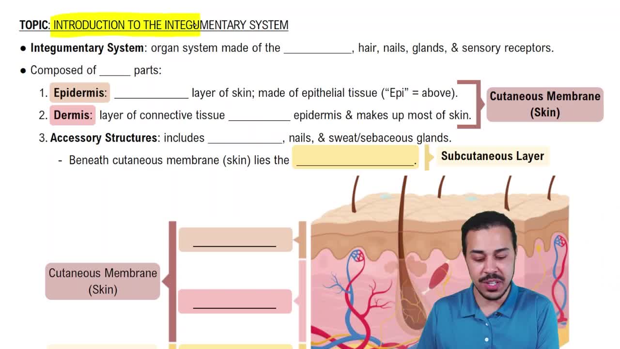
The skin, hair, nails, sweat glands, and sebaceous glands.
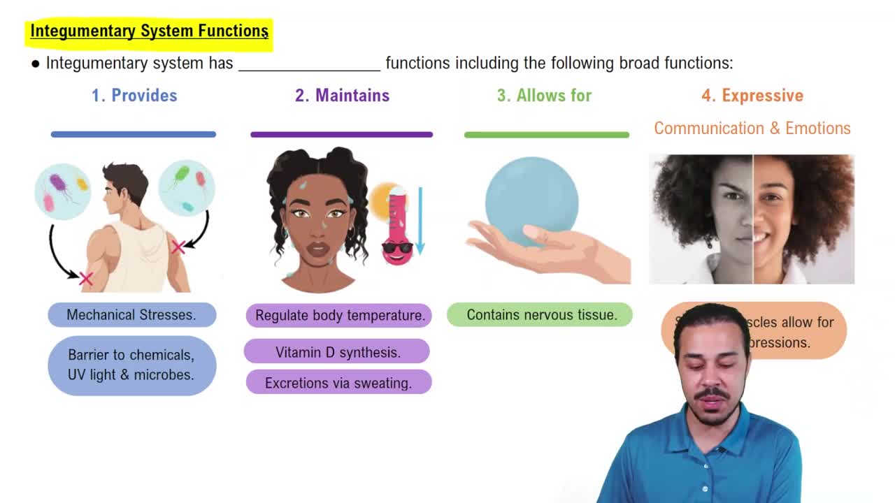
Protection, temperature regulation, sensation, excretion, vitamin D synthesis, and waterproofing.
Keratinized stratified squamous epithelium.
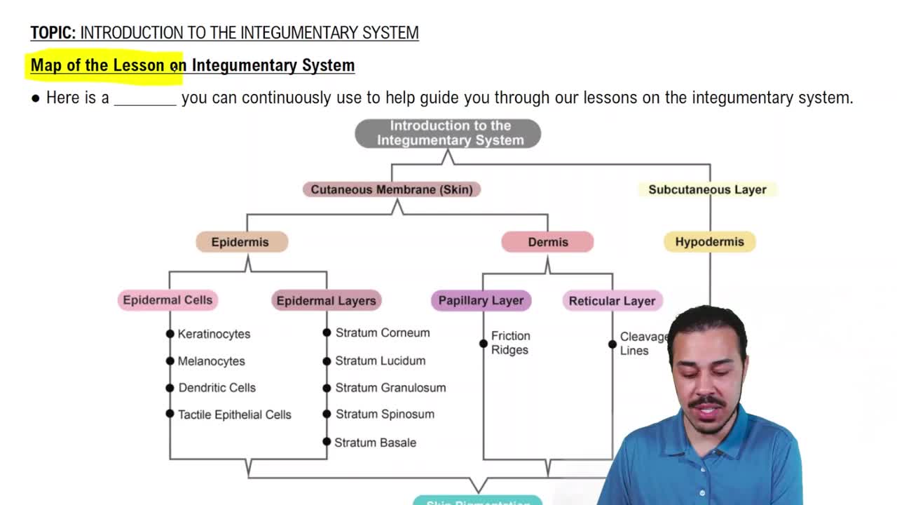
Keratocytes produce keratin; melanocytes produce pigment; Langerhans cells provide immune defense; Merkel cells function in touch sensation.
Stratum basale, stratum spinosum, stratum granulosum, stratum lucidum (only in thick skin), stratum corneum.
Basale: mitotic cells; Spinosum: desmosomes; Granulosum: keratohyalin granules; Lucidum: clear dead cells; Corneum: dead keratinized cells.
The dermis lies directly beneath the epidermis.
Contains fibroblasts, macrophages, mast cells, blood vessels, nerves, hair follicles, and glands.
Papillary layer: loose connective tissue with dermal papillae; Reticular layer: dense irregular connective tissue providing strength.
Papillary: capillaries and sensory neurons; Reticular: sweat glands, sebaceous glands, blood vessels, pressure receptors.
Includes friction ridges (fingerprints), flexion lines, and freckles.
The hypodermis lies beneath the dermis, connecting skin to underlying tissues.
Composed mainly of adipose tissue; provides insulation, energy storage, and cushioning.
Melanin, carotene, and hemoglobin.
Melanin: produced by melanocytes, brown/black pigment; Carotene: from diet, orange-yellow; Hemoglobin: red from blood in vessels.
Cyanosis: blue, low oxygen; Jaundice: yellow, liver issues; Erythema: redness, inflammation; Pallor: paleness, anemia.
Inflammation: clot formation; Organization: granulation tissue forms; Regeneration: epithelial growth and remodeling.
Hair: protection and sensation; Nails: protect digits; Sweat glands: thermoregulation; Sebaceous glands: lubrication.
Palms, soles, lips, nipples, and parts of external genitalia.
What is hair made of?
Made of keratinized dead cells arranged in a shaft.
Root: embedded in follicle; Shaft: visible part above skin.
Includes bulb (growth zone), papilla (blood supply), and sheaths (protective layers).
Lanugo: fetal hair; Vellus: fine body hair; Terminal: thick hair on scalp, eyebrows, and pubic areas.
Anagen: active growth; Catagen: regression; Telogen: resting phase.
Androgenic alopecia: genetic; Telogen effluvium: stress-related shedding.
Eccrine glands: widespread, thermoregulation; Apocrine glands: in axillary/genital areas, scent glands.
Eccrine: all over body; Apocrine: armpits, groin, around nipples.
Sebaceous glands: secrete oily sebum; Sweat glands: secrete watery sweat.
Holocrine glands that release sebum by cell rupture.
Basal cell carcinoma: most common, slow-growing; Squamous cell carcinoma: arises from keratinocytes; Melanoma: from melanocytes, most dangerous.
Loss of fluid, risk of infection, and impaired temperature regulation.
Assessed by depth (degree) and total body surface area affected.
First-degree: epidermis only; Second-degree: epidermis and part of dermis.
Destroy epidermis and dermis, may affect hypodermis; painless due to nerve damage; require grafting.