Skip to main content
Anatomy & Physiology
My Course
Learn
Exam Prep
AI Tutor
Study Guides
Textbook Solutions
Flashcards
Explore
Try the app
My Course
Learn
Exam Prep
AI Tutor
Study Guides
Textbook Solutions
Flashcards
Explore
Try the app
Back
Peripheral and Autonomic Nervous System Study Guide
You can tap to flip the card.
Connective tissue coverings of a nerve
You can tap to flip the card.
👆
Connective tissue coverings of a nerve
The three layers are
endoneurium
(around individual axons),
perineurium
(around fascicles), and
epineurium
(around the entire nerve).
Track progress
Control buttons has been changed to "navigation" mode.
1/30
Recommended videos
1:06
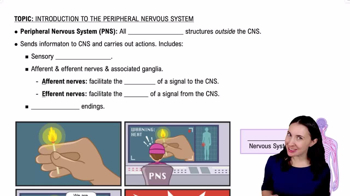
Introduction to the Peripheral Nervous System Example 1
9059
views
214
rank
3:33
Peripheral Nervous System
13005
views
332
rank
1
comments
Terms in this set (30)
Hide definitions
Connective tissue coverings of a nerve
The three layers are
endoneurium
(around individual axons),
perineurium
(around fascicles), and
epineurium
(around the entire nerve).
Main nerve plexuses
The cervical, brachial, lumbar, and sacral plexuses are networks of nerves that supply limbs and body regions.
Is there a thoracic plexus?
No, because thoracic spinal nerves mostly form intercostal nerves instead of plexuses.
Purpose of nerve plexuses
Plexuses allow nerves to redistribute fibers so that each limb muscle receives input from multiple spinal nerves, providing redundancy.
Wrist drop and associated nerve
Wrist drop is caused by damage to the
radial nerve
, leading to inability to extend the wrist.
Nerve associated with carpal tunnel syndrome
The
median nerve
is compressed in carpal tunnel syndrome, causing numbness and weakness in the hand.
Pathway of vision and decussation site
Visual signals travel from retina to optic nerve; decussation occurs at the
optic chiasm
where fibers cross.
Dorsal vs. ventral rami
Dorsal rami carry sensory and motor fibers to the back; ventral rami serve the limbs and anterior trunk.
Difference between rami and roots
Roots are the initial spinal nerve branches (dorsal root sensory, ventral root motor); rami are branches after the spinal nerve forms.
Fibers contained in dorsal and ventral rami
Both dorsal and ventral rami contain mixed fibers: sensory and motor neurons.
Functions of cranial nerves
Cranial nerves can be sensory, motor, or mixed, controlling senses, muscles, and autonomic functions.
Meaning of mixed cranial nerves
Mixed nerves carry both sensory and motor fibers.
Cranial nerves carrying somatic motor neurons
Several cranial nerves carry somatic motor neurons, including III, IV, VI, XI, and XII.
Cranial nerves carrying parasympathetic motor neurons
Cranial nerves III, VII, IX, and X carry parasympathetic motor fibers.
Bell’s Palsy and associated nerve
Bell’s Palsy is facial paralysis caused by dysfunction of the
facial nerve (cranial nerve VII)
.
Role of the phrenic nerve
The phrenic nerve controls the diaphragm and is essential for breathing.
Funny bone nerve
The 'funny bone' sensation is caused by hitting the
ulnar nerve
at the elbow.
Afferent vs. efferent neurons
Afferent neurons
carry sensory information to the CNS;
efferent neurons
carry motor commands away from the CNS.
Sympathetic division slogan
"Fight or flight" describes the sympathetic nervous system's role in stress responses.
Parasympathetic division slogan
"Rest and digest" describes the parasympathetic nervous system's role in conserving energy and digestion.
Sympathetic vs. parasympathetic fiber length
Sympathetic has short preganglionic and long postganglionic fibers; parasympathetic has long preganglionic and short postganglionic fibers.
Neurotransmitters in sympathetic division
Sympathetic preganglionic neurons release
acetylcholine
; postganglionic neurons release
norepinephrine
.
Neurotransmitters in parasympathetic division
Both preganglionic and postganglionic parasympathetic neurons release
acetylcholine
.
Neurotransmitter in somatic nervous system
Somatic motor neurons release
acetylcholine
to stimulate skeletal muscles.
Effects of sympathetic division on heart, pupils, and respiration
Increases heart rate, dilates pupils, and increases respiratory rate.
Effects of parasympathetic division on heart, pupils, and respiration
Decreases heart rate, constricts pupils, and decreases respiratory rate.
Location of sympathetic preganglionic fibers
Sympathetic preganglionic fibers originate in the thoracolumbar region of the spinal cord (T1-L2).
Somatic vs. autonomic neuron chains
Somatic division uses a single neuron from CNS to muscle; autonomic division uses a two-neuron chain (preganglionic and postganglionic).
Parasympathetic vs. sympathetic innervation
Parasympathetic innervates mainly visceral organs for rest functions; sympathetic innervates organs for fight or flight responses.
Craniosacral vs. thoracolumbar divisions
Parasympathetic division is craniosacral; sympathetic division is thoracolumbar.
 BackBack
 1:06
1:06