Skip to main content
Anatomy & Physiology
My Course
Learn
Exam Prep
AI Tutor
Study Guides
Textbook Solutions
Flashcards
Explore
Try the app
My Course
Learn
Exam Prep
AI Tutor
Study Guides
Textbook Solutions
Flashcards
Explore
Try the app
Back
Set #3 Anatomy & Physiology: Ribs, Spine, & Upper Extremities
You can tap to flip the card.
Nasal septum
You can tap to flip the card.
👆
Nasal septum
The vertical midline partition dividing the nasal cavity into right and left halves, made of the vomer, perpendicular plate, and cartilage.
Track progress
Control buttons has been changed to "navigation" mode.
1/35
Recommended videos
Guided course
6:29
Spine (Vertebral Column)
10183
views
216
rank
Guided course
5:17
The Spine Example 1
7235
views
149
rank
Terms in this set (35)
Hide definitions
Nasal septum
The vertical midline partition dividing the nasal cavity into right and left halves, made of the vomer, perpendicular plate, and cartilage.
Zygomatic arch
The bony cheek arch formed by the fusion of the temporal process of the zygomatic bone and the zygomatic process of the temporal bone.
Bony orbit
The cone-shaped skeletal cavity enclosing the eye and associated muscles, formed by 7 different skull bones.
Lambdoid suture
The arched line of articulation across the posterior skull, connecting the occipital bone with the paired parietal bones.
Sagittal suture
The midline articulation line on the top of the skull, connecting the right and left parietal bones.
Coronal suture
The line of articulation running crown-like across the top of the skull, connecting the frontal bone to the parietal bones.
Squamous suture
The lower lateral articulation line on each side of the skull, connecting the temporal bone to the parietal bone.
Frontal suture
A suture line separating the two halves of the frontal bone during skull development, usually fusing in early childhood.
Frontonasal suture
The horizontal line of articulation across the root of the nose, connecting the frontal bone with the nasal bones.
Intermaxillary suture
The midline articulation line connecting the right and left maxilla bones directly under the nose.
Anterior (frontal) fontanel
The largest diamond-shaped soft spot in an infant skull, located at the junction of the sagittal, coronal, and frontal sutures.
Posterior (occipital) fontanel
A small, triangular soft spot in an infant skull, located posteriorly at the junction of the sagittal and lambdoid sutures.
Sphenoidal fontanel
An irregular soft spot in an infant skull located anterolaterally at the junction of the sphenoid, parietal, temporal, and frontal bones.
Mastoid fontanel
An irregular soft spot in an infant skull located posterolaterally at the junction of the temporal, parietal, and occipital bones.
Hyoid bone
A U-shaped bone in the mid-neck above the larynx; unique because it does not articulate directly with any other bone.
Costal cartilage
Segments of hyaline cartilage connecting the ribs to the sternum, providing flexibility to the rib cage.
Ribs
12 pairs of curved flat bones forming the lateral walls of the thoracic cage; right vs. left side orientation must be determined.
Sternal end of rib
The blunt, flattened anterior end of the rib that attaches to the costal cartilage.
Vertebral end of rib
The posterior end of the rib featuring the head, neck, and tubercle; articulates with the thoracic vertebrae.
Costal tubercle
A small knob-like projection on the posterior surface of the rib near the head; articulates with the transverse process of a thoracic vertebra.
Costal groove
A shallow trench along the internal inferior border of a rib; protects intercostal nerves and blood vessels.
True ribs
Rib pairs 1 through 7; attach directly to the sternum via individual costal cartilages.
False ribs
Rib pairs 8 through 12; do not attach directly to the sternum (8-10 fuse to cartilage of rib 7).
Floating ribs
Rib pairs 11 and 12; a subcategory of false ribs with no anterior attachment to the sternum or other cartilage.
Sternum
The breastbone; a flat bone located along the anterior midline of the thoracic cage.
Manubrium
The superior, widest, and triangular portion of the sternum.
Jugular notch
A shallow, central depression on the top edge of the manubrium of the sternum.
Body of sternum
The long, central, middle section of the sternum.
Xiphoid process
The small, inferior, pointed cartilaginous projection at the lower tip of the sternum.
Cervical region of spine
The neck region of the spine, consisting of 7 vertebrae (C1−C7).
Thoracic region of spine
The chest region of the spine, consisting of 12 vertebrae (T1−T12) that articulate with ribs.
Lumbar region of spine
The lower back region of the spine, consisting of 5 large, stout vertebrae (L1−L5).
Sacral region of spine
The pelvic region of the spine, consisting of 5 fused vertebrae forming the sacrum.
Coccygeal region of spine
The tailbone region at the bottom of the spine, consisting of 3 to 5 small fused vertebrae.
Vertebral body (centrum)
The thick, disc-shaped anterior weight-bearing section of a vertebra.
 BackBack
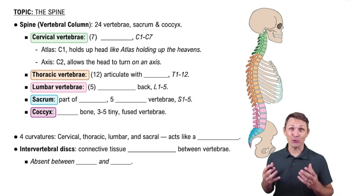
 6:29
6:29