 Back
BackSpinal Cord and Reflexes - Anatomy & Physiology

 0:41
0:41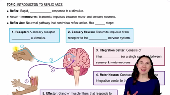

Terms in this set (83)
Rapid, automatic nerve responses controlled by the spinal cord only, not the brain.
Approximately 18 inches long and ½ inch wide.
At the L1–L2 vertebral level.
The spinal cord is bilaterally symmetrical.
Posterior median sulcus.
Anterior median fissure, which is deeper than the posterior sulcus.
Cervical enlargement.
Lumbar enlargement.
Conus medullaris.
Cauda equina (horse tail nerves).
Filum terminale.
31 spinal cord segments.
In the dorsal root ganglion.
Dorsal roots carry sensory information.
Ventral roots carry motor information.
Mixed nerves containing both sensory and motor fibers.
By the fusion of dorsal (sensory) and ventral (motor) roots.
Protect the spinal cord and carry blood supply.
Inflammation of the meninges.
Dura mater (outer), arachnoid mater (middle), and pia mater (inner).
Dense connective tissue.
Fat and blood vessels.
In the epidural space.
Subarachnoid space.
Cerebrospinal fluid (CSF).
Shock absorber and provides nutrients.
The innermost meningeal layer attached directly to the spinal cord.
Denticulate ligaments.
Axons, located superficially.
Neuron cell bodies.
Processes sensory information.
Controls motor neurons.
Controls autonomic neurons.
Carry sensory information to the brain.
Carry motor commands from the brain.
Epineurium.
Perineurium.
Endoneurium.
Back muscles and skin.
Limbs and body wall.
An area of skin supplied by one spinal nerve.
Used to locate nerve damage.
Varicella-zoster virus.
Affects sensory nerves causing a painful rash.
Only ventral rami form plexuses.
C1–C5.
Phrenic nerve from the cervical plexus.
C5–T1.
Motor and sensory control of the upper limb.
Median nerve.
Controls extensor muscles of the arm and forearm.
Controls many hand muscles.
T12–L4.
Femoral nerve.
L4–S4.
Sciatic nerve, from the sacral plexus.
Afferent fibers.
Efferent fibers.
Receptor activation.
Sensory neuron activation.
Interneuron processing in CNS.
Motor neuron activation.
Effector response (muscle or gland action).
Reflexes that are present at birth.
Reflexes that are learned through experience.
Skeletal muscle responses.
Organ functions.
A reflex with one synapse, the fastest type.
A reflex with multiple synapses.
In the spinal cord.
In the brain.
Maintains posture by responding to muscle stretch.
Muscle spindle.
Prevents muscle damage by inhibiting excessive tension.
Pulls a body part away from painful stimuli.
Flexors contract while extensors relax during a reflex.
The opposite leg stabilizes the body during withdrawal reflex.
Occurs on the same side of the body.
Occurs on the opposite side of the body.
Yes, the brain can enhance or inhibit reflexes.
Increases the strength of a reflex.
Decreases the strength of a reflex.
Normal in infants but abnormal in adults, indicates CNS damage if present.