 Indietro
Anatomy & Physiology: Blood - Structure, Function, and Disorders
 2:36
2:36
Termini in questo insieme (29)
Transport: delivers O2, nutrients, wastes, hormones.
Regulation: maintains temperature, pH, fluid volume.
Protection: prevents blood loss and infection.
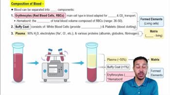
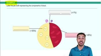
Blood is connective tissue with plasma (nonliving fluid matrix) and formed elements (RBCs, WBCs, platelets).
Percentage of blood volume occupied by RBCs; normal ~45% (males 47% ± 5%, females 42% ± 5%).
Sticky, opaque fluid with metallic taste; color varies with O2 content; pH 7.35–7.45; ~8% body weight; volume 4-6 L.
About 90% water with dissolved solutes including nutrients, gases, hormones, wastes, and plasma proteins like albumin, globulins, and fibrinogen.
Biconcave, anucleate cells filled with hemoglobin; flexible due to spectrin protein; diameter ~7.5 μm.
Protein with 4 polypeptide chains (2 alpha, 2 beta), each bound to a heme group with iron that binds O2.
Transport respiratory gases: O2 binds to heme forming oxyhemoglobin; CO2 binds to globin forming carbaminohemoglobin.
Formation of RBCs in red bone marrow from hematopoietic stem cells, regulated by erythropoietin (EPO).
Released by kidneys in response to hypoxia; stimulates RBC production; inhibited by high O2 or RBC count.
Amino acids, iron (stored as ferritin), vitamin B12, and folic acid are essential for erythropoiesis.
Live 100-120 days; old RBCs are phagocytized in spleen; hemoglobin is recycled.
Condition with low O2-carrying capacity due to blood loss, decreased RBC production, or increased RBC destruction.
Genetic disorder with mutated hemoglobin S causing crescent-shaped RBCs that rupture easily and block vessels.
Complete cells with nuclei; defend against disease; classified as granulocytes or agranulocytes.
Include neutrophils, eosinophils, basophils; contain cytoplasmic granules; phagocytic.
Most abundant WBCs (50-70%); multilobed nucleus; kill bacteria via respiratory burst.
2-4% of WBCs; bilobed nucleus; combat parasitic worms; involved in allergies.
Rarest WBCs (0.5-1%); contain histamine which promotes inflammation and attracts other WBCs.
Include lymphocytes and monocytes; lack visible granules; important in immunity and phagocytosis.
25% of WBCs; large nucleus; T cells attack infected cells; B cells produce antibodies.
3-8% of WBCs; largest WBC; differentiate into macrophages; phagocytic and activate lymphocytes.
Cell fragments from megakaryocytes; contain clotting chemicals; form platelet plugs to stop bleeding.
1. Vascular spasm (vasoconstriction)
2. Platelet plug formation
3. Coagulation (fibrin clot formation)
Intrinsic pathway triggered by blood factors; extrinsic pathway triggered by tissue factor; both activate factor X.
Thrombin converts fibrinogen to fibrin; fibrin mesh stabilizes clot; factor XIII cross-links fibrin.
Plasma proteins numbered I to XIII; vitamin K required for synthesis of some factors.
Based on presence of A and/or B antigens on RBCs; antibodies in plasma react against absent antigens.
Rh– mother exposed to Rh+ fetus blood can form antibodies causing hemolytic disease in subsequent Rh+ pregnancies; prevented by RhoGAM.