 Indietro
Anatomy & Physiology: Cardio, Vasculature, and Respiratory System
 3:06

Termini in questo insieme (21)
Pulmonary circuit: Right side receives O2 poor blood from the body and pumps it to the lungs.
Systemic circuit: Left side receives O2 rich blood from lungs and pumps it throughout the body.
Epicardium: visceral layer of serous pericardium. Myocardium: cardiac muscle arranged in circular and spiral patterns.
Endocardium: endothelium resting on connective tissue lining internal heart walls.
Intercalated discs join adjacent cardiac muscle cells; contain fasciae adherens and gap junctions that allow electrical impulses to spread for synchronized contraction.
AV valves open when atrial pressure > ventricular pressure, allowing blood flow into ventricles.
They close when ventricular pressure > atrial pressure, preventing backflow (LUB sound).
Dense connective tissue surrounding valves; anchors valve cusps, prevents valve dilation, serves as insertion for cardiac muscle, and blocks direct electrical impulse spread between atria and ventricles.
Impulse starts at SA node → spreads to atria and AV node → pauses at AV node → travels via Bundle of His → divides into left and right bundle branches → Purkinje fibers spread impulse to ventricles.
Elastic arteries: largest, near heart, high elastin content. Muscular arteries: medium-sized, thick tunica media, active in vasoconstriction.
Arterioles: smallest, regulate blood flow into capillaries.
Tunica intima: innermost, endothelium.
Tunica media: smooth muscle and elastin.
Tunica externa: connective tissue outer layer.
Continuous: least permeable, tight junctions.
Fenestrated: pores, more permeable.
Sinusoids: large gaps, highly permeable, found in liver, bone marrow, spleen.
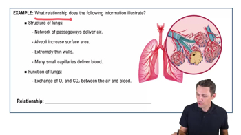
Reduces surface tension in alveoli to prevent collapse during exhalation, essential for newborn breathing.
Pulmonary ventilation, external respiration, transport of respiratory gases, and internal respiration.
Double-layered sac:
parietal pleura lines thoracic cavity;
visceral pleura covers lungs; pleural cavity contains lubricating fluid to reduce friction during breathing.
Quiet inhalation: diaphragm and external intercostals.
Forced inhalation: sternocleidomastoid, scalenes, pectoralis minor, serratus posterior superior, erector spinae.
Located posteriorly in trachea; contracts to decrease tracheal diameter during coughing and sneezing to expel air forcefully.
Right ventricle pumps O2 poor blood into pulmonary trunk → pulmonary arteries → lungs for gas exchange → pulmonary veins carry O2 rich blood to left atrium.
Remnant of fetal foramen ovale; allowed blood to bypass lungs in fetus; closes after birth.
Brachiocephalic trunk (right common carotid and right subclavian), left common carotid artery, and left subclavian artery.
Regulate blood flow into capillary beds by contracting or relaxing to control tissue perfusion.
Superior vena cava, inferior vena cava, and coronary sinus.
Venous blood from digestive organs passes through liver capillaries before returning to systemic circulation, allowing nutrient processing and detoxification.
Provides collateral blood flow to the brain by connecting anterior and posterior cerebral circulations, ensuring brain perfusion if one artery is blocked.